ENERGY DEVICE FRONTIER

この記事の要点
シミ治療は、機器の名前からではなく、色素が表皮のおよそ0.2ミリのどの深さにあるのかを読むことから始まります。この記事では、シミを深さで「表皮型」「混在型」「真皮型」の3つのタイプに分け、タイプごとの治療の考え方を、17の症例とあわせてご紹介します。もう一つの鍵は、肌の司令塔である線維芽細胞の状態です。働きが落ちた「低活動モード」の細胞は元に戻せるため、当院はCrossTALK® protocolで線維芽細胞を再生モードへ戻してからエネルギー治療を行う設計を採り、その有効性と安全性を国際誌に報告しています。自分のシミのタイプと、肌の状態。この二つを知ることが、再発しにくい肌への近道です。
治療の適応、回数、ダウンタイム、費用、感じ方には個人差があります。治療内容は診察のうえで決定します。
表皮の厚さは、およそ0.2ミリです。 同じ茶色に見えるシミでも、色素がこの0.2ミリより浅くあるのか、深くあるのかで、選ぶべき治療は別のものになります。浅いシミに合う照射が、深いシミには届かない。深いシミに合う照射を浅いシミに重ねれば、肌への負担だけが残ることもあります。
シミ治療の情報は溢れています。機器の名前で治療を選べるように見える時代だからこそ、私が診察で最初にしているのは、機器を選ぶことではありません。この肌の、どの深さで、何が起きているのかを読むことです。
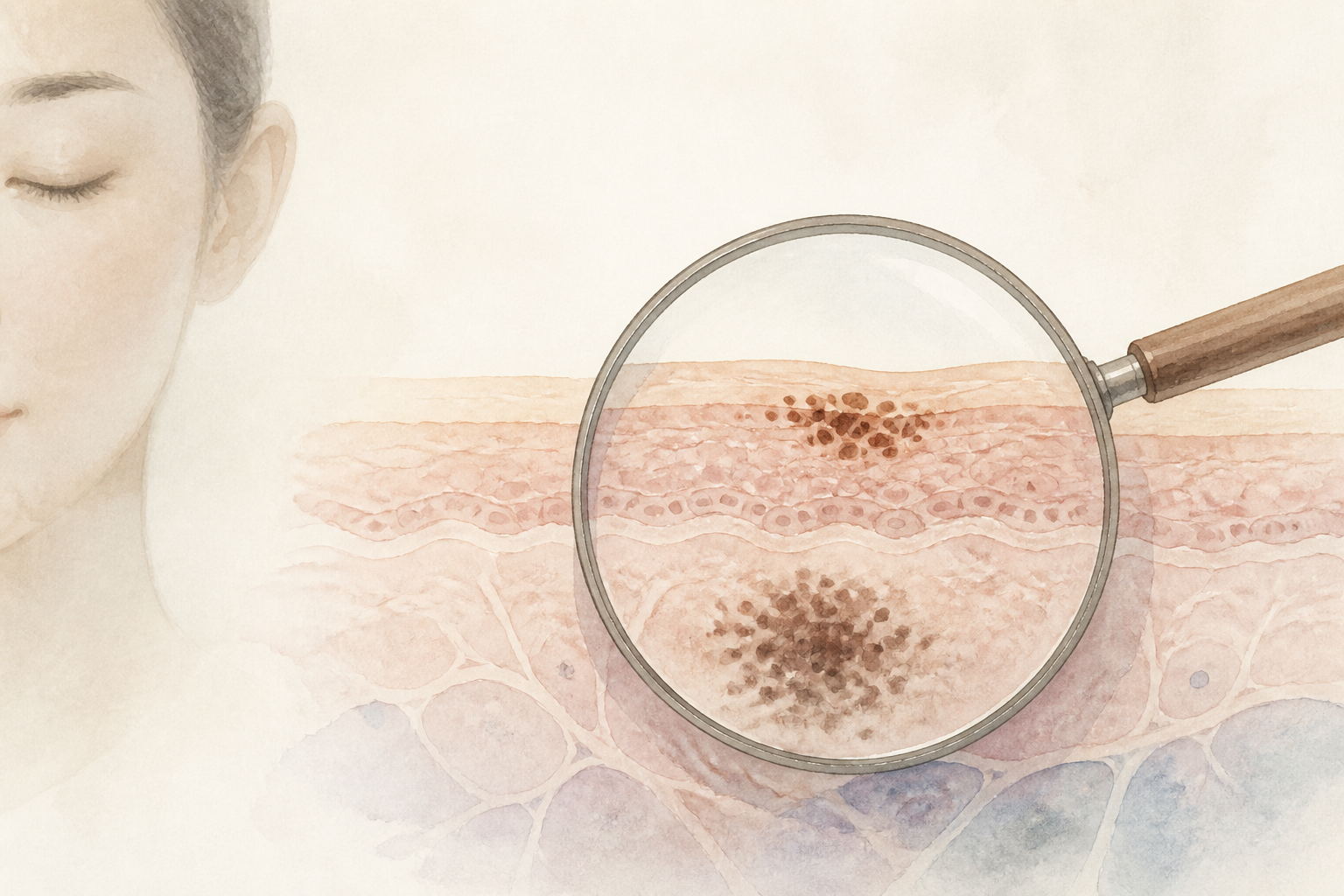
見た目が似ていても、シミは同じではありません
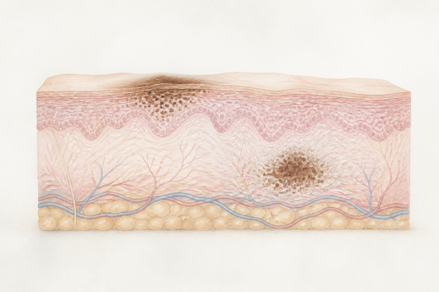
輪郭のはっきりした日光性色素斑。細かなそばかす。左右対称に広がる肝斑。真皮にあるADM。炎症の後に残った色素沈着。
診察では色素の種類だけでなく、赤み、乾燥、炎症傾向、摩擦の癖、紫外線を浴びる機会、これまでの治療歴まで確認します。照射に対する肌の反応は、色素そのものと同じくらい、その周りの肌環境で変わるからです。
だから、すぐに照射をしない、という判断があります。目立つ一点を急いで消すことが、その方の5年後の肌にとって最善とは限りません。当てない判断まで含めて、シミ治療です。
あなたのシミは、どのタイプでしょうか
診察で最初に読み取るのは、色素の深さです。色素がおよそ0.2ミリの上にあるのか、上下に重なっているのか、下が中心なのか。この記事では、それを3つのタイプに分けてお話しします。
Type Map — 0.2mm
あなたのシミは、どのタイプ?
日光性色素斑・脂漏性角化症・雀卵斑 など
日光性色素斑や雀卵斑+肝斑の併存 など
ADM・太田母斑 など
TYPE 01 表皮型は、色素が0.2ミリの上だけにあるシミです。日光性色素斑、脂漏性角化症、雀卵斑(そばかす)がここに入ります。TYPE 02 混在型は、これらの表皮のシミに、左右対称に広がる肝斑が重なっているタイプ。実際の診察室で最も多く出会うのが、この混在型です。TYPE 03 真皮型は、色素の中心が0.2ミリより下にあるタイプで、ADM(後天性真皮メラノサイトーシス)や太田母斑が代表です。
鏡の前での、見分けの目安
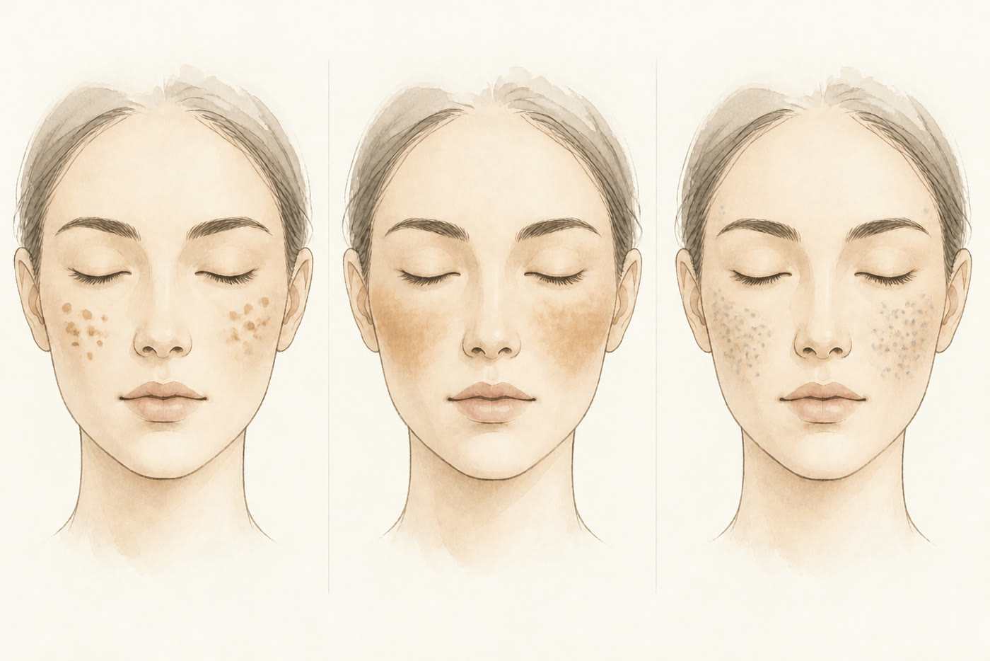
左から、表皮型・混在型・真皮型のイメージ。
輪郭のはっきりした茶色が、ぽつり、ぽつりと独立して見える —— 表皮型の傾向。
輪郭のぼやけた色が、頬骨の上に左右ほぼ対称に広がって見える —— 肝斑をともなう混在型の傾向。
茶色というより、灰色や青みがかって見える —— 真皮型の傾向。
タイプは、見た目だけでは正確に判定できません。診察で、シミの種類・深さ・肌環境を合わせて確定します。なお、ほくろ(色素性母斑)は「シミ」とは分けて考えます。診断を優先し、治療をするかどうかは、そのうえで判断します。
シミは、雑草に似ています
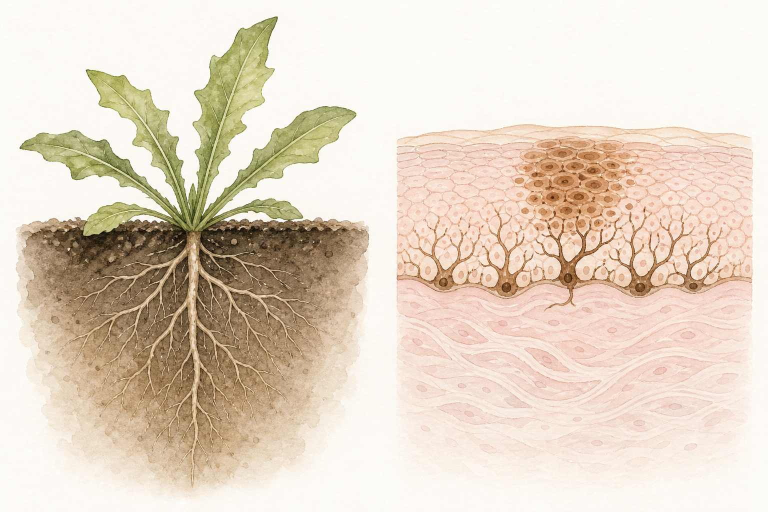
地面の上に見えている葉を刈れば、その日のうちに景色は変わります。けれど土の中に根が残っていれば、同じ場所からまた生えてきます。シミにも、これとよく似た構造があります。
見えている茶色は、表皮に溜まったメラニン(肌の色を作る色素)です。雑草でいえば、地上に出た葉。0.2ミリの上にあるこの部分は、レーザーで壊すことができます。
根にあたるのは、表皮の底で色素を作り続けるメラノサイト(色素を作る細胞)の過剰な働きと、それを刺激し続ける肌環境です。紫外線、摩擦、炎症、ホルモンの変化。根が働き続けている限り、表面の色素を消しても、時間とともに同じ場所へ色が戻ってきやすくなります。
「取れたはずのシミが、また出てきた」の多くは、葉だけを刈って、根がそのまま残っている状態です。3つのタイプは、この比喩でも読めます。葉だけが茂っているのが表皮型。葉の下で根が働き続けているのが混在型。根が深いところにあるのが真皮型です。
TYPE 01|表皮型 —— 0.2ミリの上だけにあるシミ
輪郭のはっきりした日光性色素斑。細かく散らばる雀卵斑。わずかに盛り上がる脂漏性角化症。いずれも色素の主座は表皮にあり、雑草でいえば、葉が茂っている状態です。このタイプの治療は、まず、見えている色素を最小限のエネルギーで壊すことから始まります。
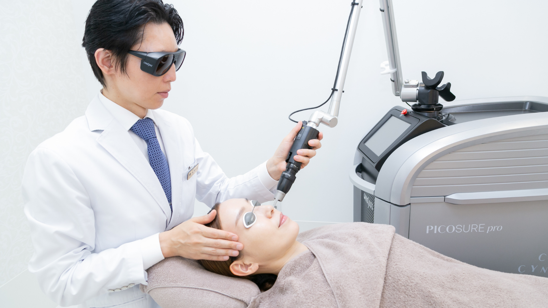
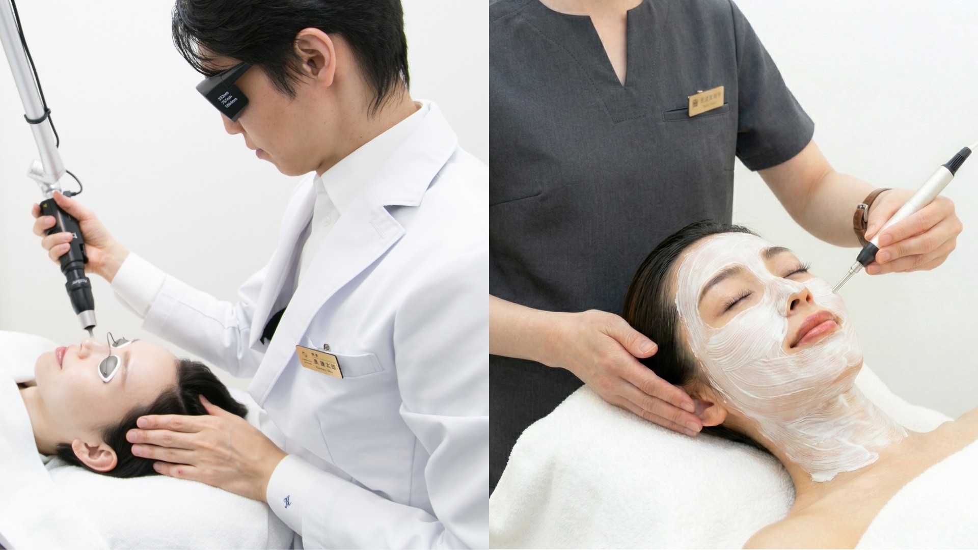
PICOSURE proは、755nmのピコ秒レーザーです。ごく短い時間でエネルギーを届ける特性を、色素へのアプローチと、肌全体の質感を考える治療に使います。
ピコレーザーの照射で最も重要なのは、出力の強さではありません。どのくらいのエネルギーなら色素だけを壊せるのか、という下限の見極めです。この境目を、閾値フルエンスと呼びます。
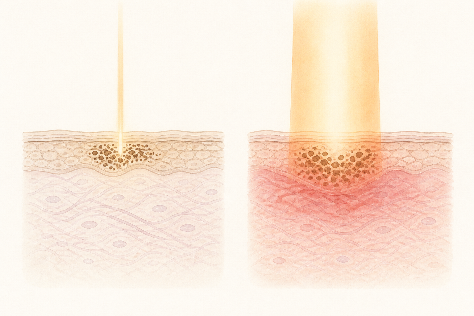
エネルギーは、高くするほど効きそうに見えます。実際には、必要以上のエネルギーは周囲の正常な組織まで反応させ、炎症後色素沈着(治療の刺激で、かえって色が濃く残ること)のリスクを高めます。壊すべきものだけを壊し、周りはしっかり守る。これが、私の考えるシミ治療の極意です。
Selective Protection
シミだけを、狙い撃つ。
25〜31%:Chan JC et al. Lasers Surg Med 2016/2.67%:奥謙太郎による自院例の集計
臨床データでは、PICOSURE proの治療後に炎症後色素沈着が生じた割合は2.67%——私自身が自院例を集計した数字です。閾値フルエンスを見極めた、必要最小限のエネルギーによる照射が、この数字を支えています。シミ治療に対して効果と安全性が高いピコレーザーであることは、基礎医学データと臨床データの両面から示されています。
シミは、背景で決まる
同じ表皮型のシミでも、その背景にある肌の状態によって、治療の設計は変わります。鍵を握るのは、真皮で働く線維芽細胞です。コラーゲンを作り、色素を作る細胞の働きをコントロールし、免疫や血管の調整にまで関わる。いわば、肌の司令塔にあたる細胞です。
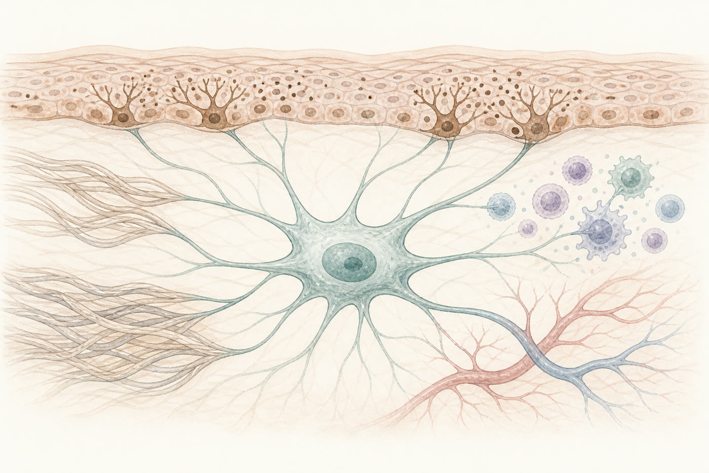
この司令塔には、状態があります。健康な線維芽細胞は、コラーゲンをつかんで、ぐっと伸びています。この物理的な「伸び」そのものが「活動しろ」というシグナルになり、コラーゲンを活発に作り続ける——再生モードです。加齢や紫外線でコラーゲンが壊れると、細胞はつかむものを失って縮こまり、眠ったように働きが落ちる——低活動モード。さらに進むと細胞分裂が止まり、炎症と分解を促す物質を出す側に回ってしまう——老化モードです(Fisher GJ et al. Arch Dermatol 2008 ほか)。
Fibroblast Status
肌の司令塔、線維芽細胞の3つのモード
若い肌の表皮型は、線維芽細胞が再生モードで働いています。エネルギーデバイスの熱は、目覚めている細胞のコラーゲン合成をさらに加速させる方向に働くため、PICOSURE proとエレクトロポレーション「デルマシオ」を組み合わせるヒルズフェイシャル®だけで、色素と質感の両方にアプローチできます。
一方、光老化が進んだ表皮型では、司令塔が低活動モードに落ちています。低活動モードの線維芽細胞は、エネルギーへの反応性そのものが下がっており、熱だけを与えられても十分には起き上がりません。それどころか、準備の整っていない肌に大きなエネルギーが加わると、熱がそのまま負担として働くことがあります。
大切なのは、老化モードと違い、低活動モードは元に戻せるということです。ここが、治療の介入ポイントになります。当院はこうした肌では、CrossTALK® protocolで先に線維芽細胞へはたらきかけます。
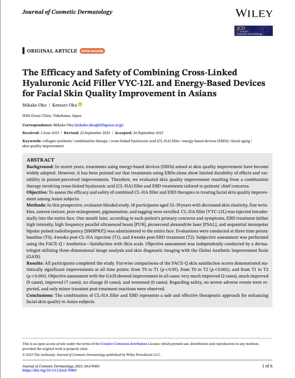
PUBLISHED IN
The Efficacy and Safety of Combining Cross-Linked Hyaluronic Acid Filler VYC-12L and Energy-Based Devices for Facial Skin Quality Improvement in Asians
架橋型ヒアルロン酸(分解されにくい構造にしたヒアルロン酸)を皮内に入れると、真皮の内圧が上がり、縮こまった線維芽細胞が物理的に伸ばされます。失われていた「つかまる足場」を作り直し、「再生を開始せよ」という合図を細胞に届ける。低活動モードの細胞が再生モードへ戻り、環境が整ってから、レーザーのエネルギーを届けます。この2段階のプロトコールは18名の患者さまで前向きに検証し、評価者判定で全員の改善と、重篤な有害事象がなかったことを国際誌に報告しています。効果・経過には個人差があります。
CASE PHOTOS — TYPE 01
表皮型の症例
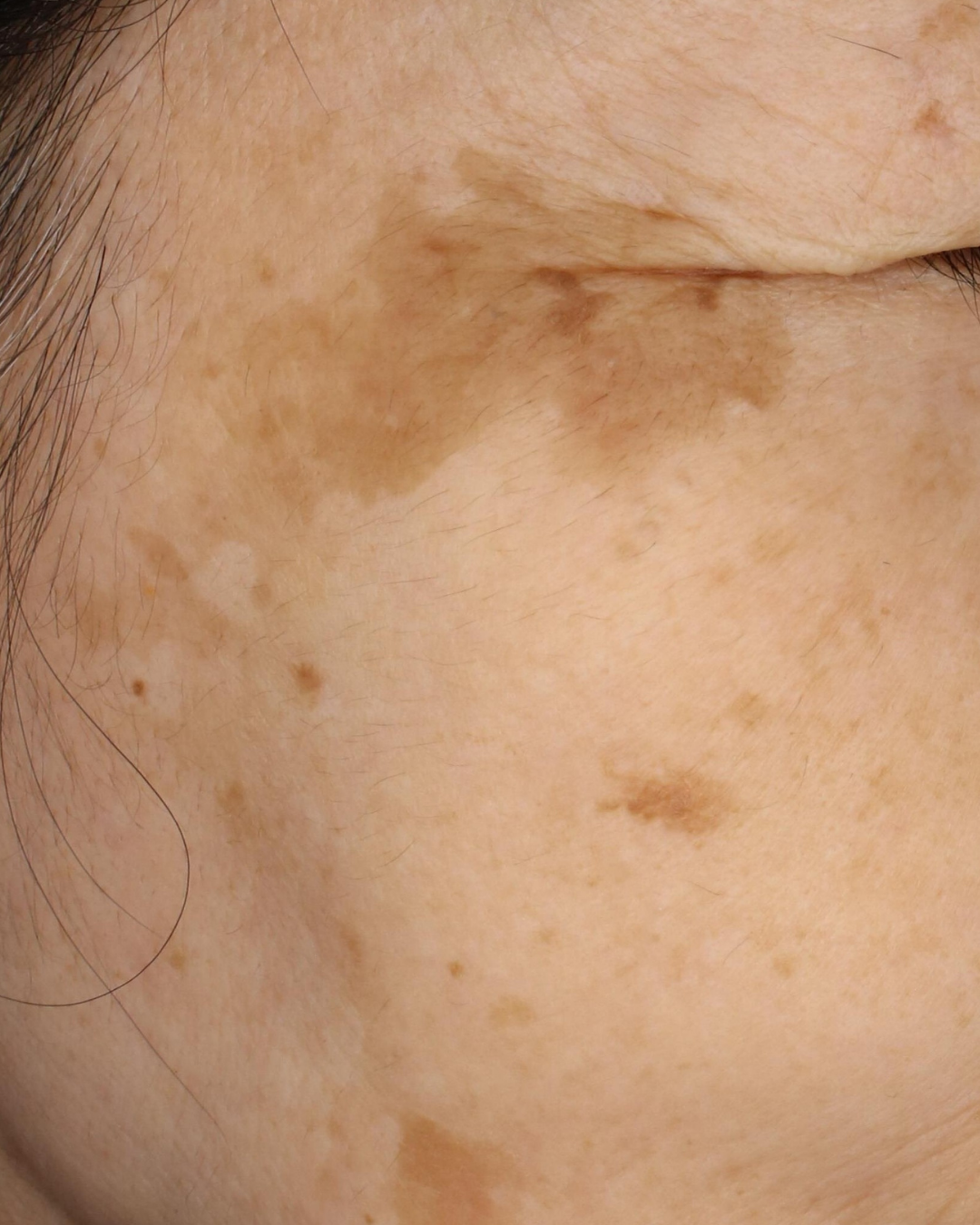
施術前
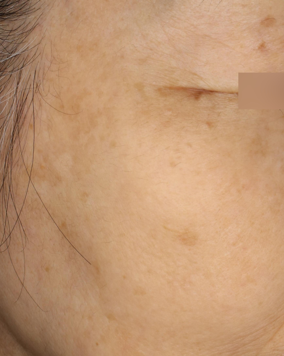
施術後
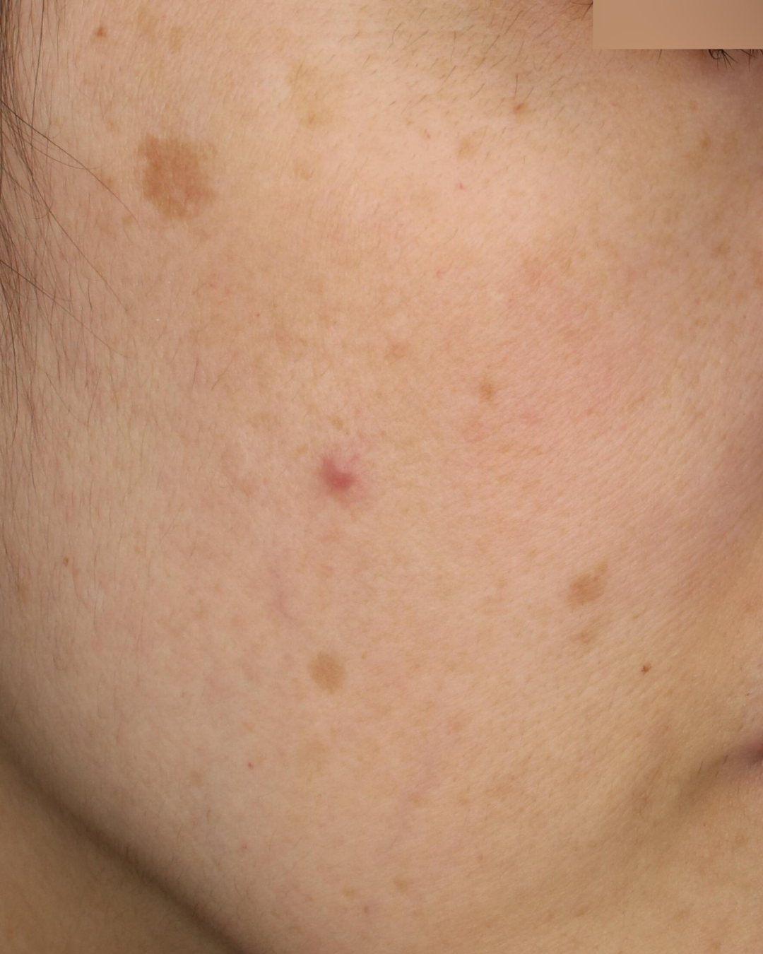
施術前
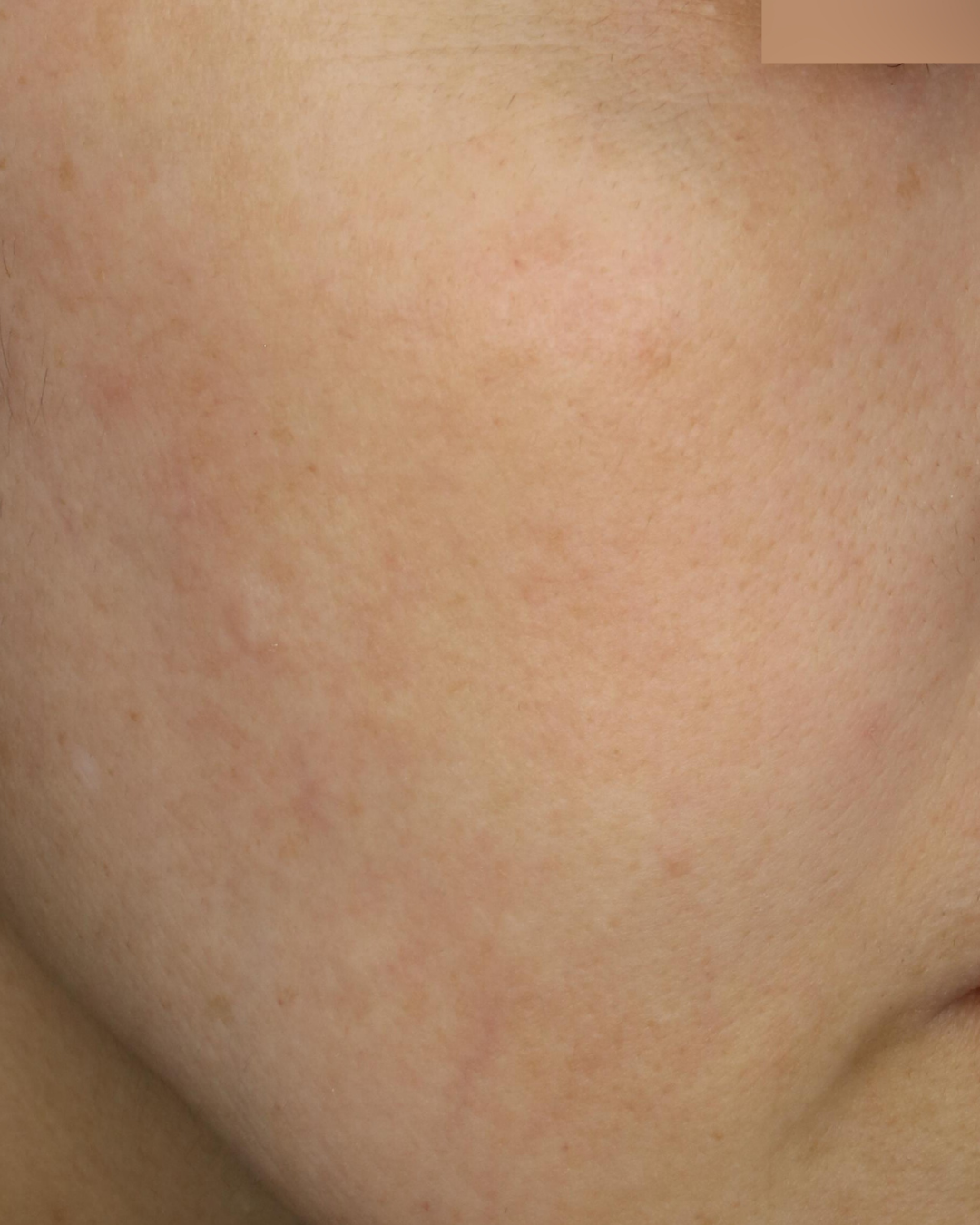
施術後
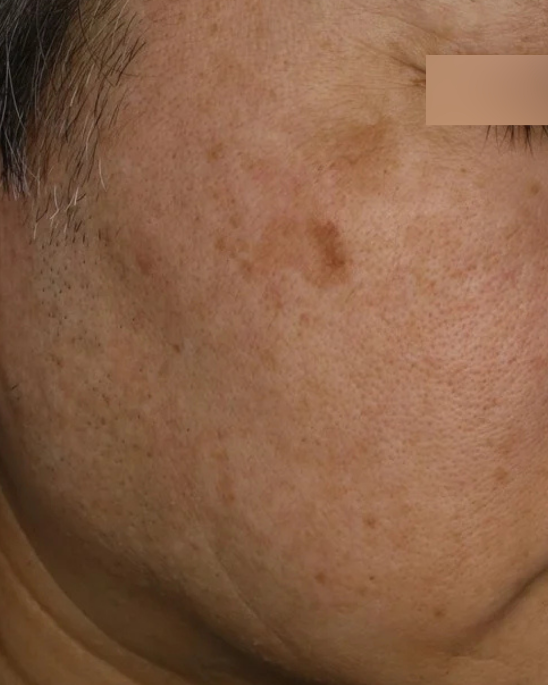
施術前
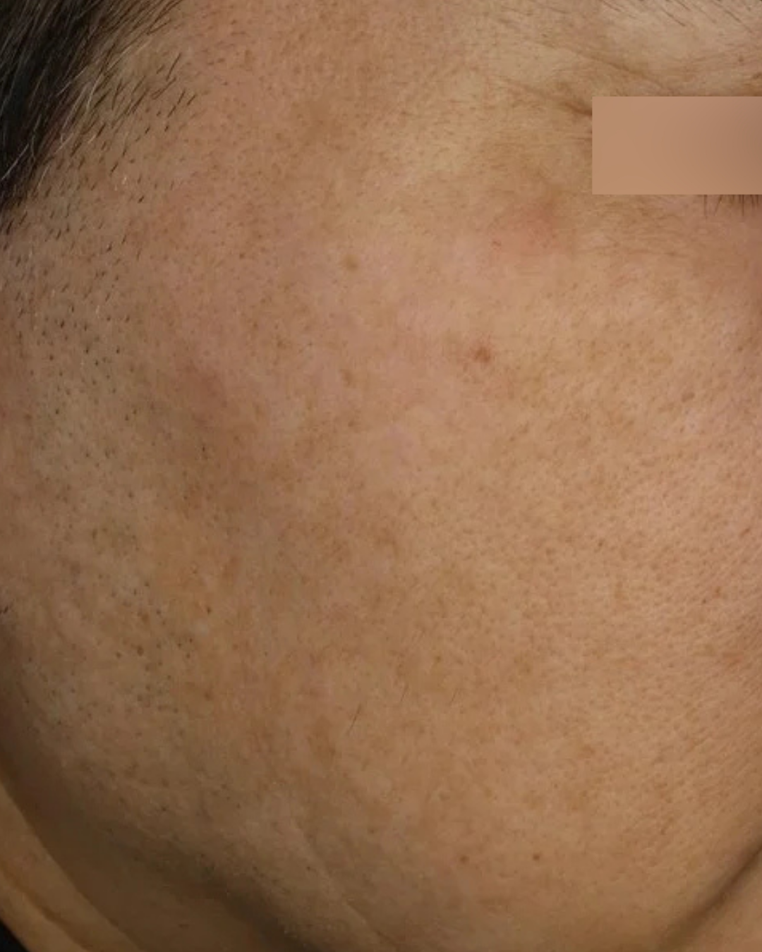
施術後
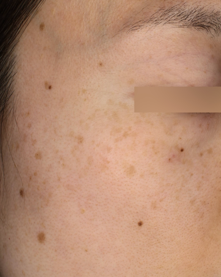
施術前
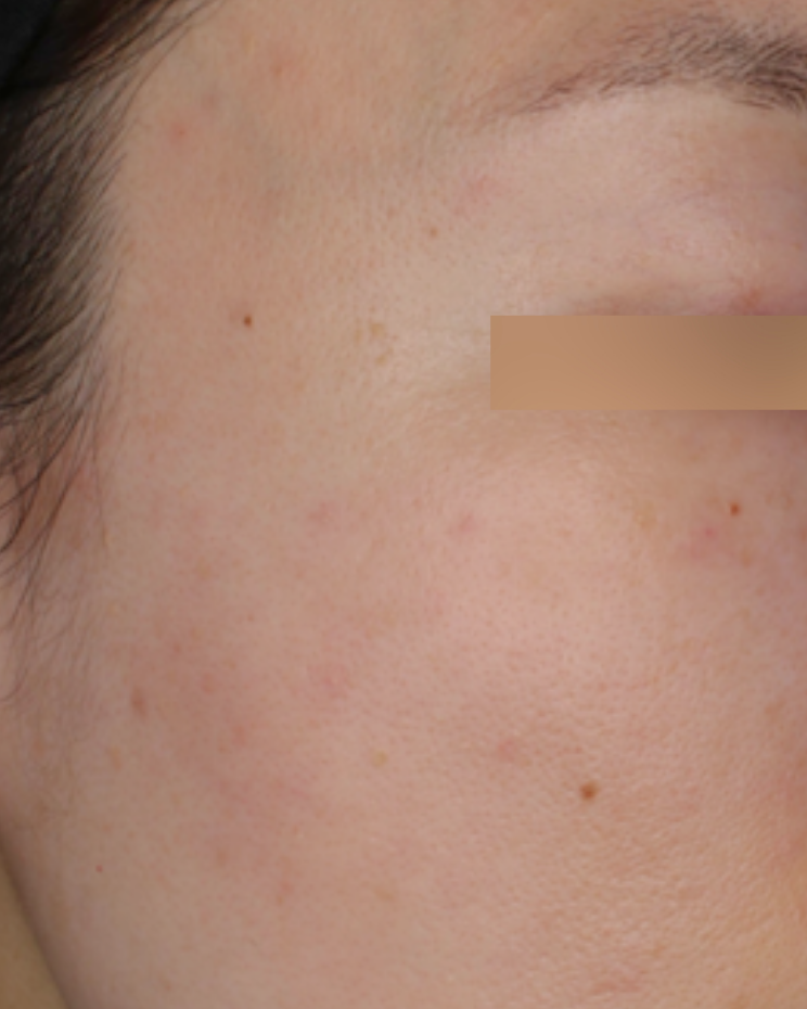
施術後
※症例写真は一例です。撮影条件、撮影時期、光の当たり方、肌の状態、治療経過には個人差があります。写真は治療効果を保証するものではなく、治療の適否は診察のうえで判断します。
TYPE 02|混在型 —— 0.2ミリの上下に、シミが重なっている
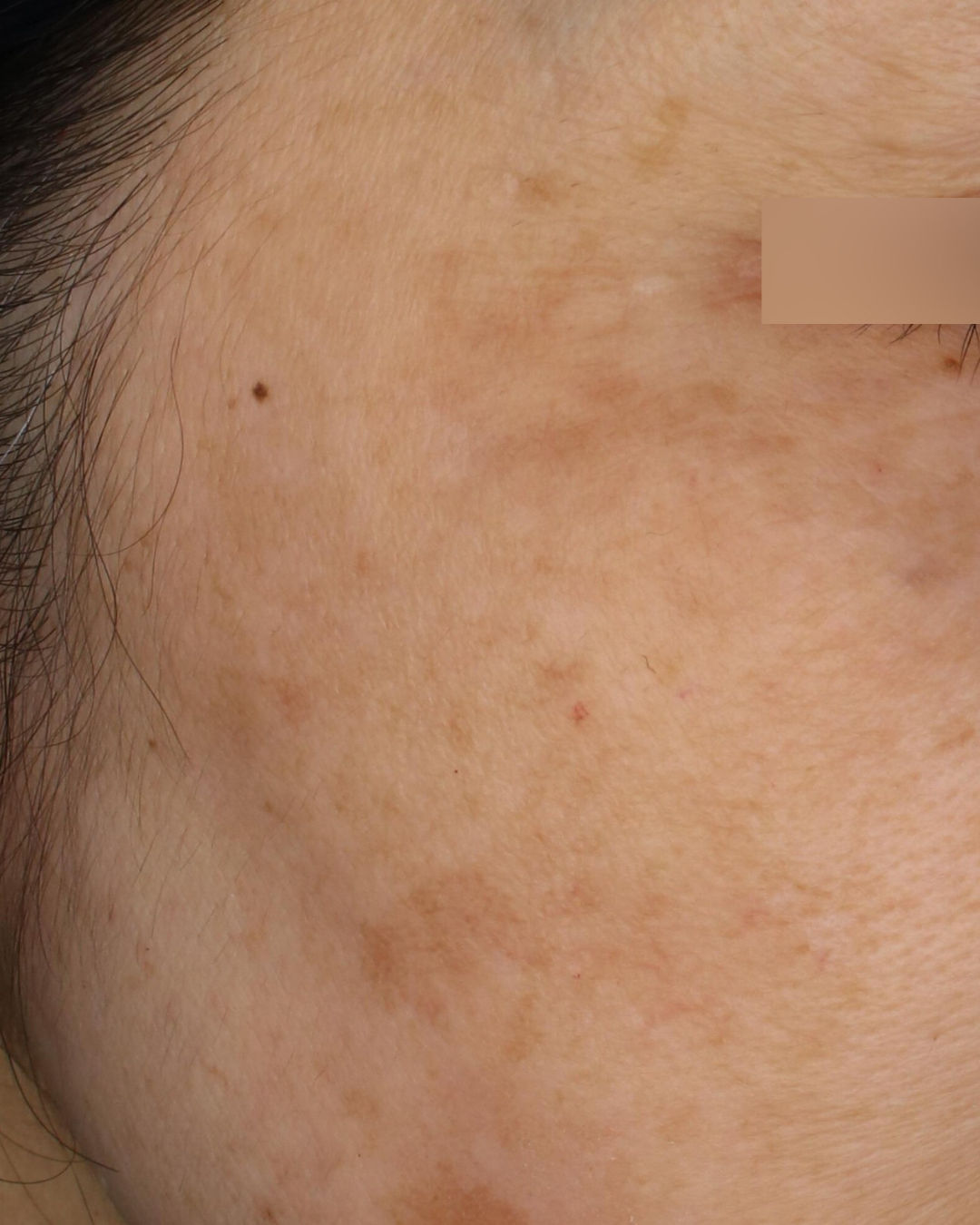
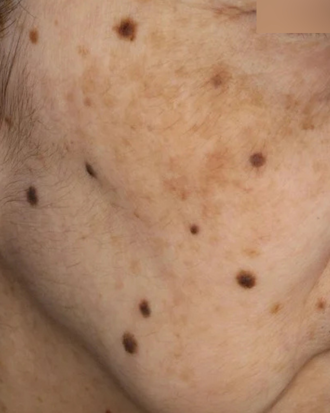
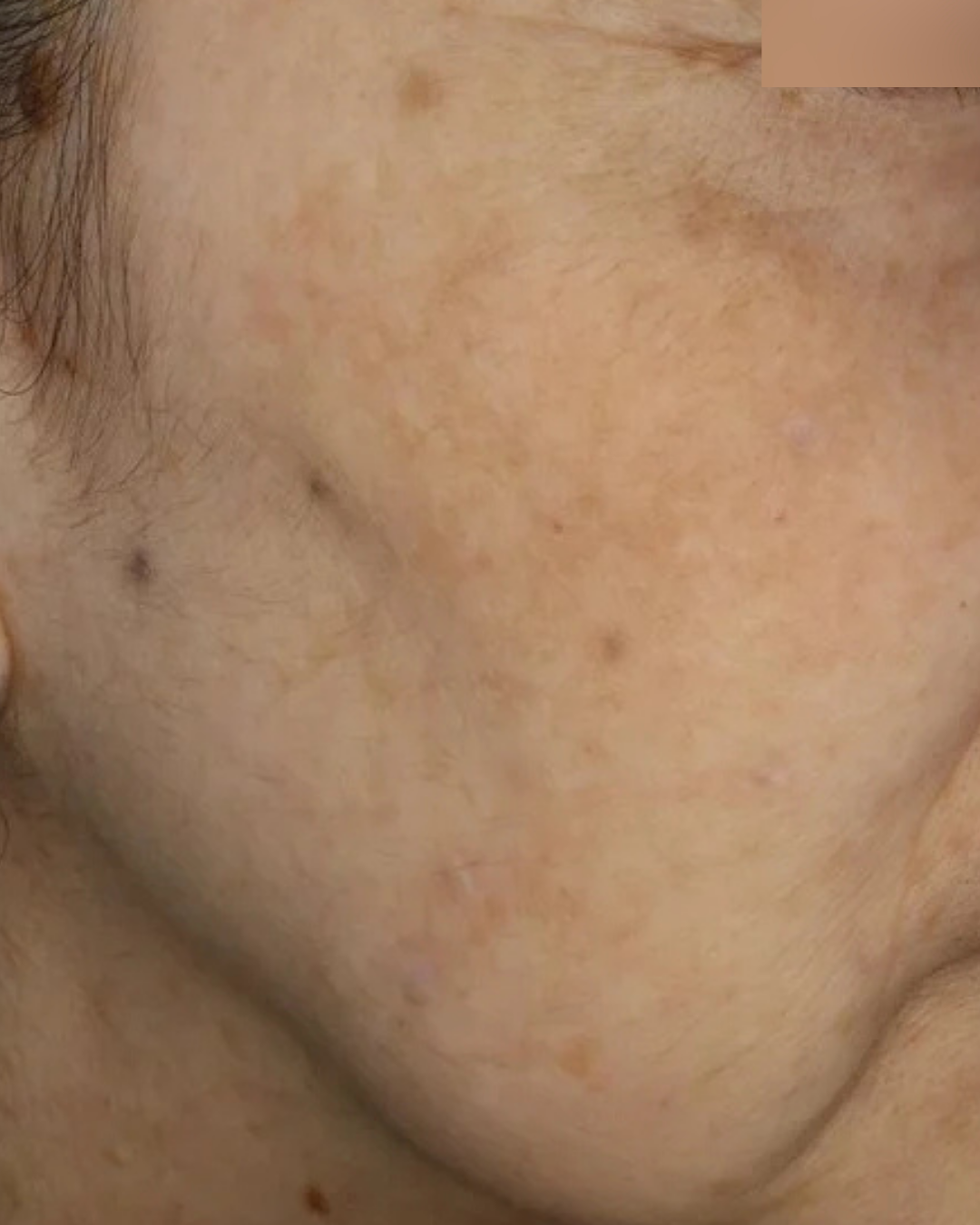
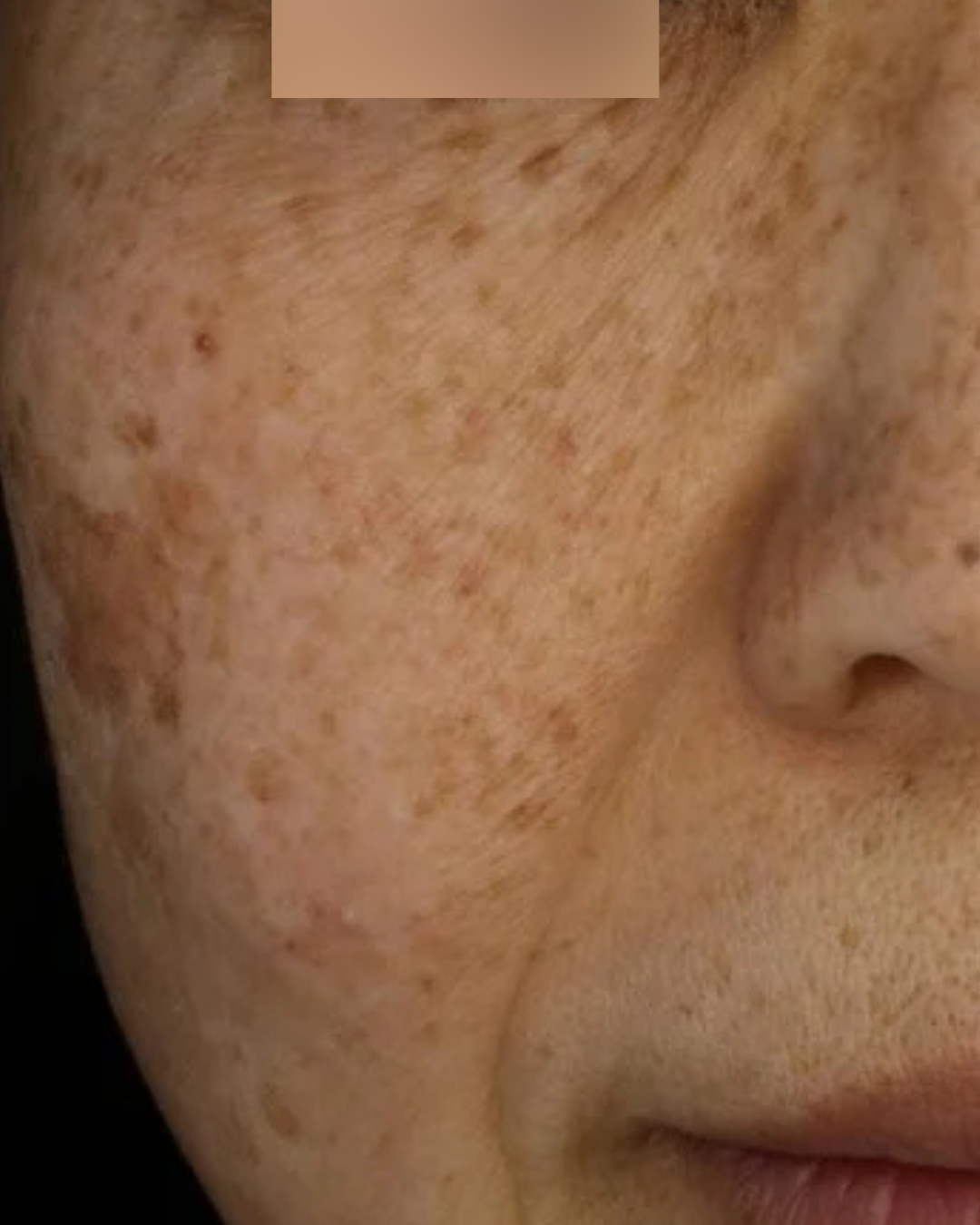
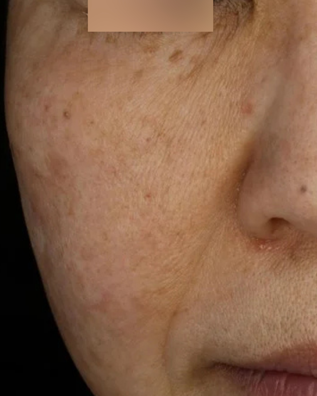
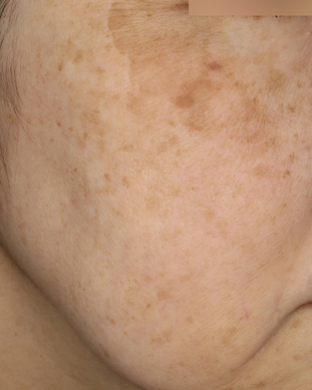
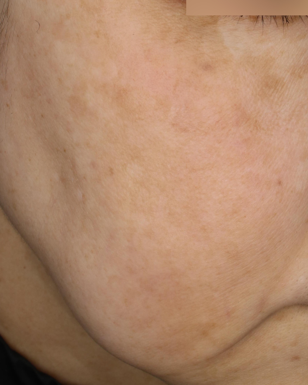
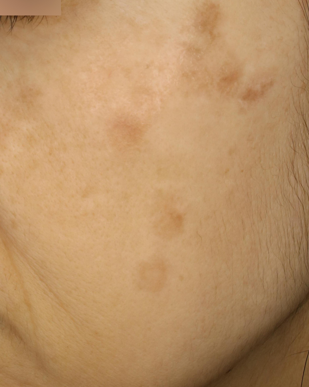
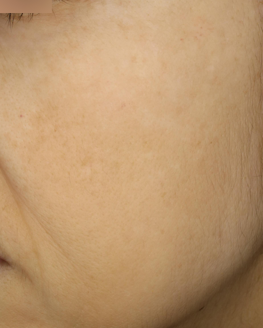
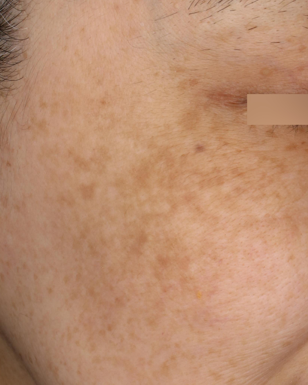
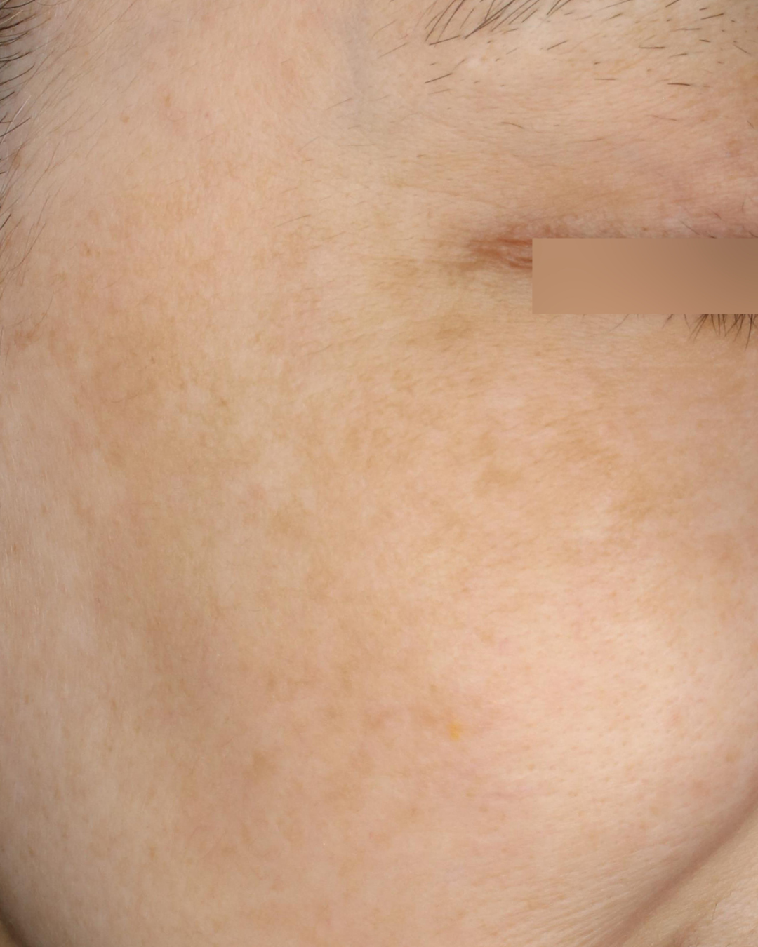
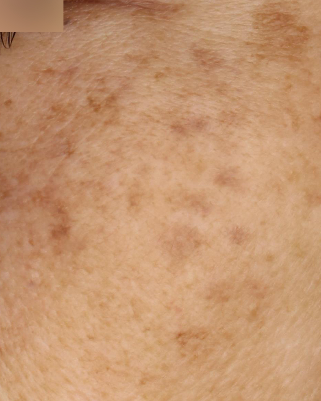
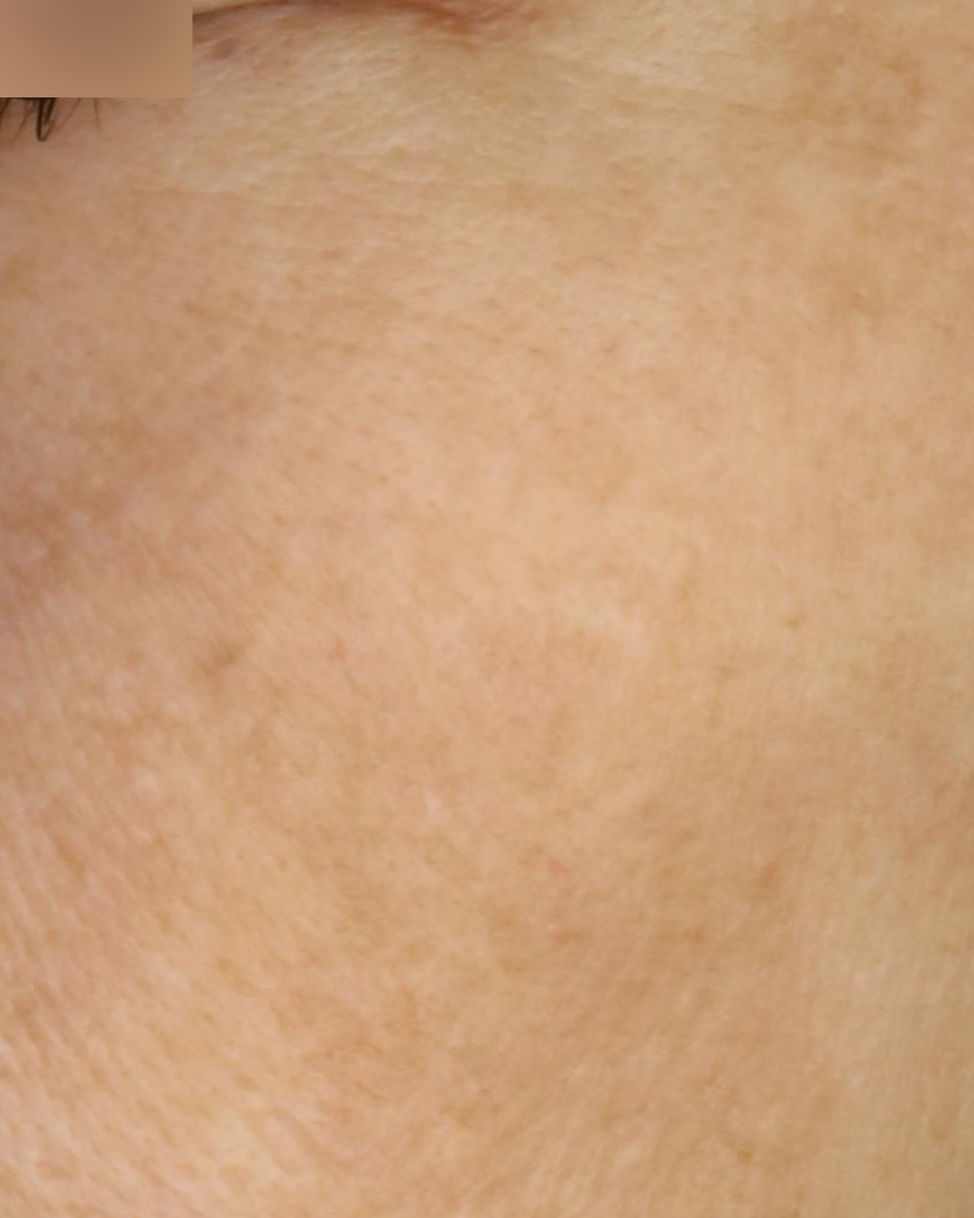
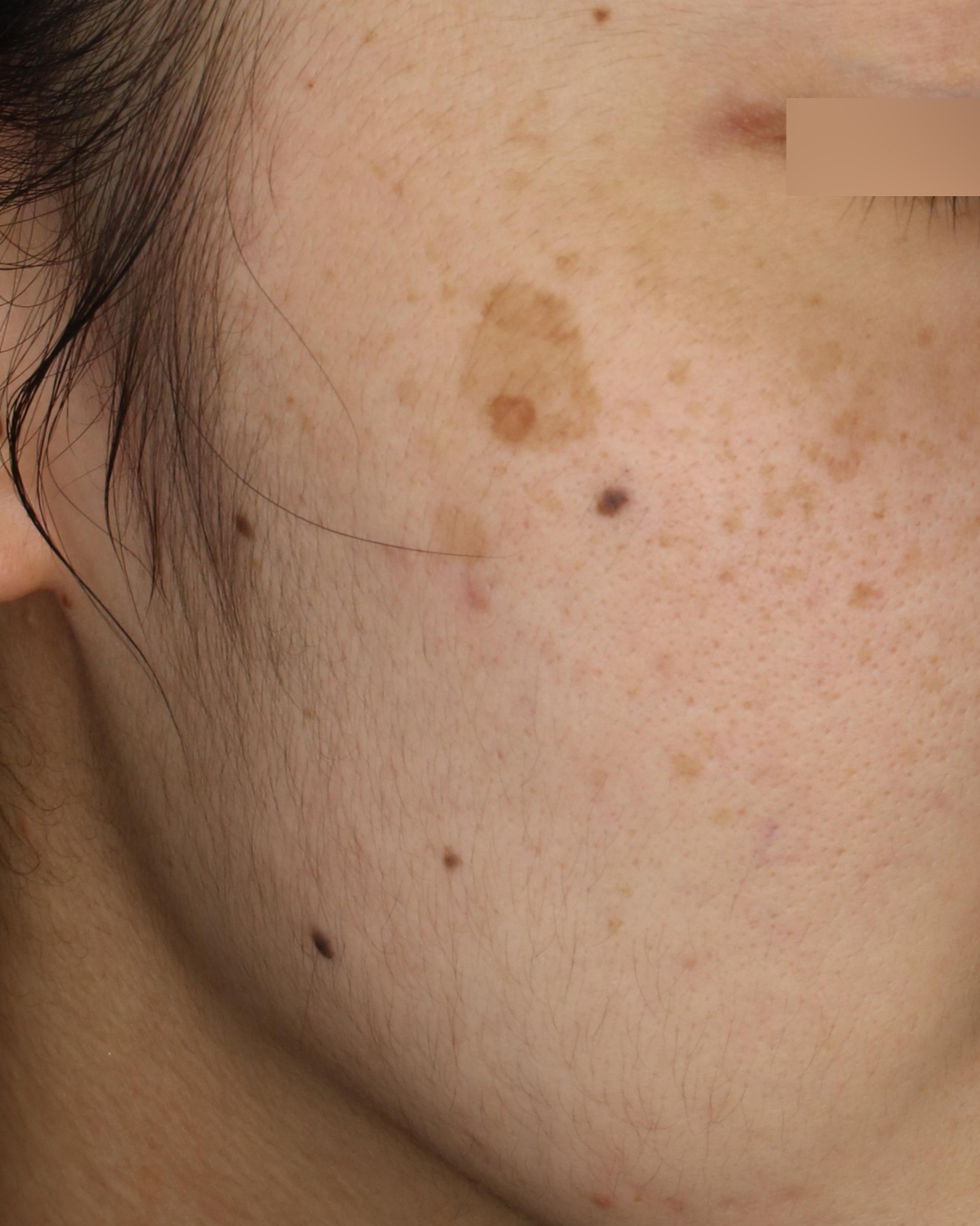
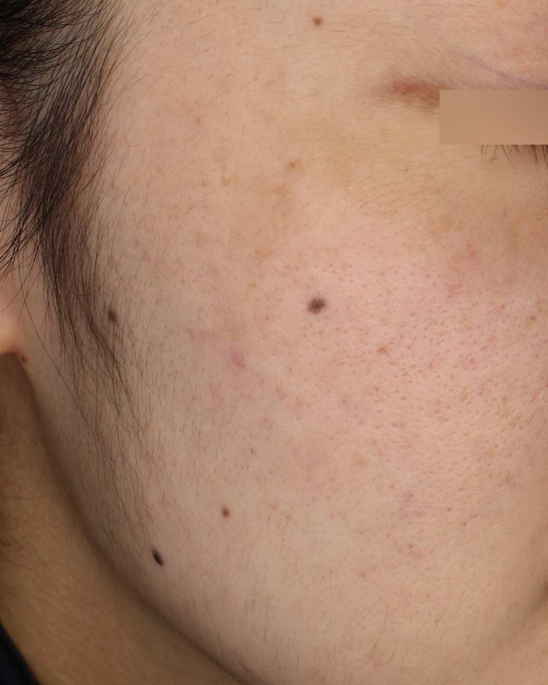
実際の診察室で最も多いのが、このタイプです。輪郭のはっきりした日光性色素斑や雀卵斑の背後に、左右対称の肝斑がうっすらと広がっている。この記事でご紹介する症例でも、半数以上が混在型にあたります。
混在型が難しいのは、表皮のシミに効く照射が、肝斑にはかえって刺激になりうることです。肝斑の根は、色素そのものより、色素を作り続けるメラノサイトの過剰な働きと、それを刺激する肌環境にあります。肝斑が体調の変化や肌への刺激で悪化や再発を起こしやすいのはこのためで、良かれと思った強い照射が、かえって根を刺激することもあります。
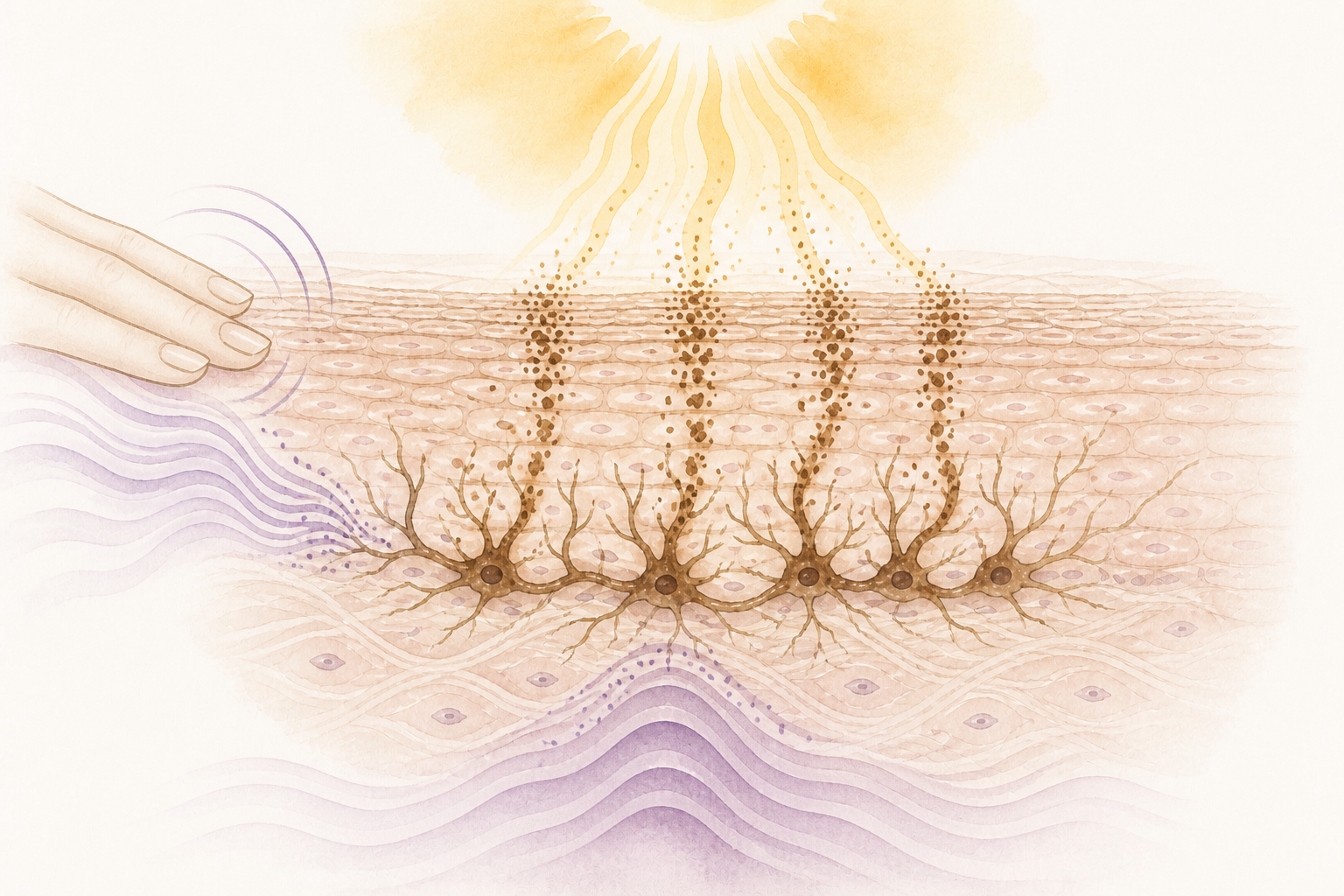
だから混在型の治療は、レーザーだけでは完結しません。刺激を抑えた照射設計に、外用・内服、紫外線対策、摩擦を減らすスキンケアを組み合わせ、根の働きそのものを鎮めていきます。葉を刈る治療と、根を弱らせる治療。この二つが揃ってはじめて、再発しにくい状態を目指せます。
家でできることも、根を弱らせる治療の一部です。可視光まで防ぐ日焼け止めを選ぶ。こすらない。外用を続ける。トラネキサム酸などの内服は、期間を決めて使う。診察では、この続け方まで含めてお伝えします。
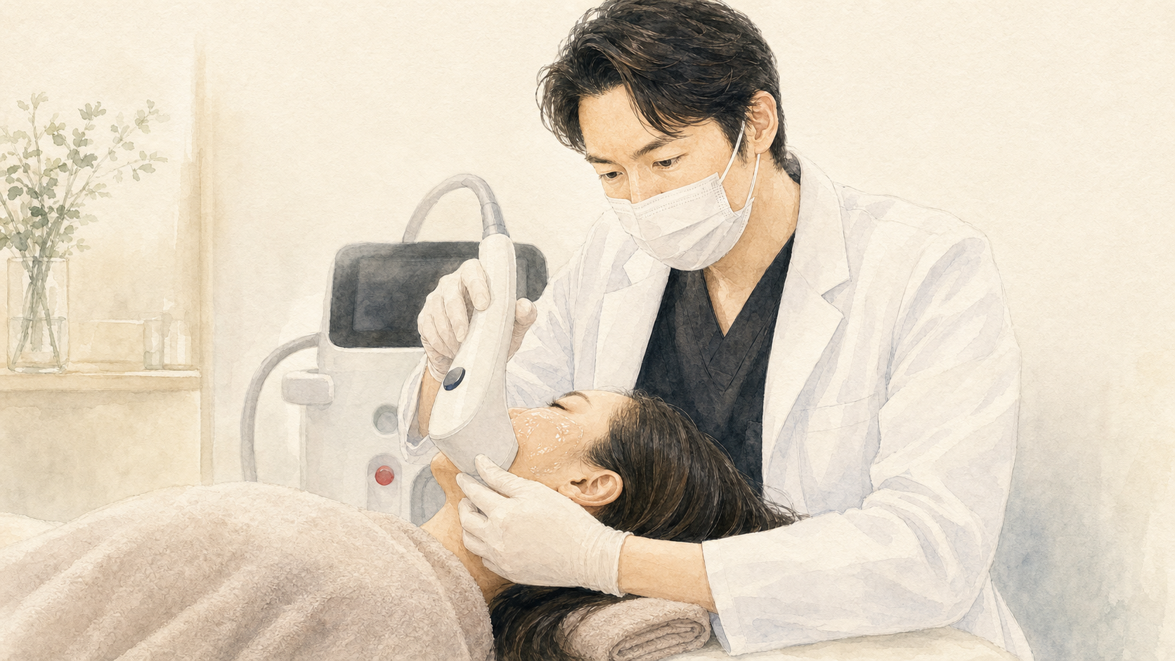
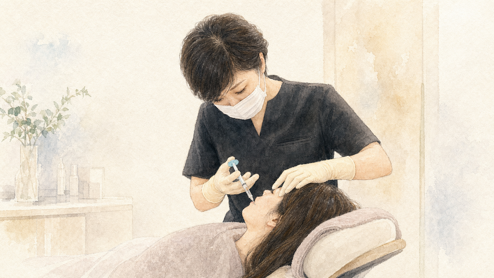
そして混在型の根に対して、当院にはもうひとつの答えがあります。肝斑の根では、老化した線維芽細胞が「色素をつくれ」という指令を出し続けていることが報告されています(Ali L, Al Niaimi F. Int J Dermatol. 2025)。表皮型の章でみた司令塔のモードでいえば、老化モードに入った細胞が声を出し続け、そのまわりに低活動モードの細胞が広がっている状態です。老化モードの細胞そのものは、もう元に戻せません。けれど、まわりの低活動モードの細胞を再生モードへ戻し、真皮の環境を立て直すことはできます。それがCrossTALK® protocolです。架橋型ヒアルロン酸で線維芽細胞がつかまれる足場を先につくり、司令塔の機能を取り戻してからエネルギーを与える。司令塔が働きを取り戻すと、色素を作る細胞への過剰な指令が鎮まる方向に働くことも示唆されています。
雑草にたとえるなら、固い土のまま葉を刈り続けるのではなく、先に土を耕してから根に向かう、という治療です。土台が整うと、一回ごとの出力を上げる必要はなくなり、刺激で悪化しやすい肝斑でも、エネルギーを負担ではなく、回復のきっかけとして使えるようになります。
CASE PHOTOS — TYPE 02
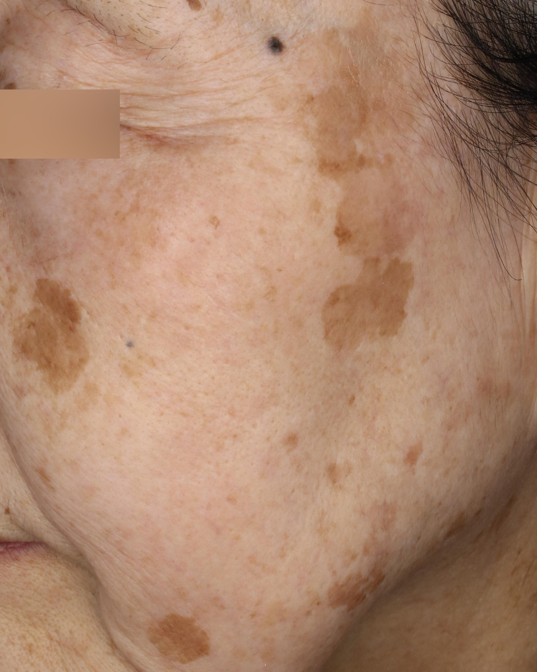
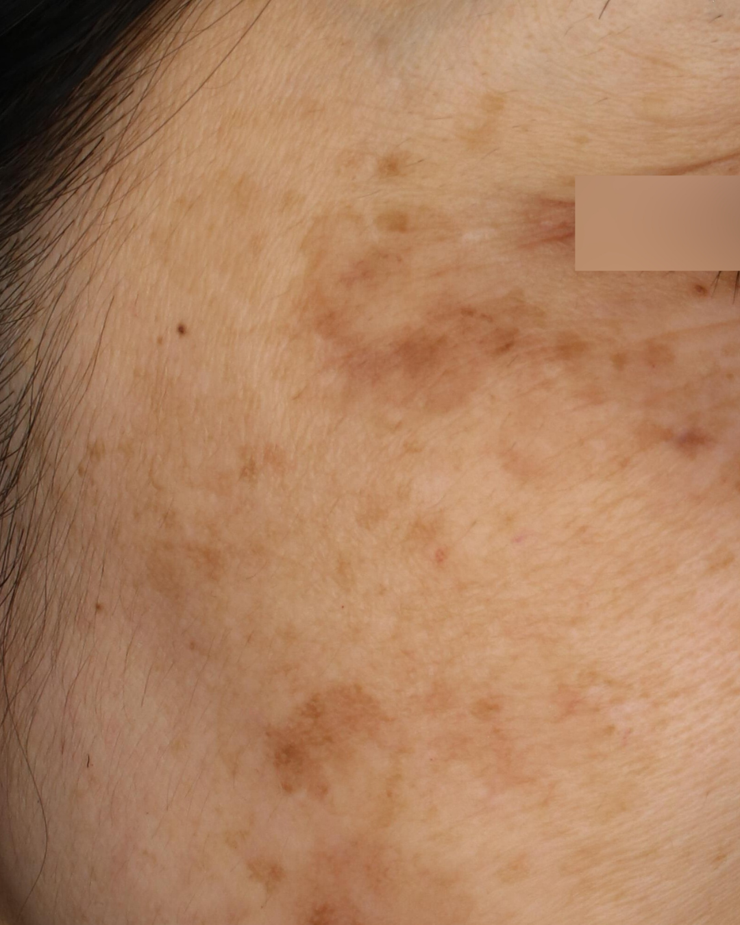
混在型の症例
肝斑を含む混在型は、症例ごとに設計が変わります。色素性母斑を含む複合的な症例も、診断のうえで治療方針を分けて対応しています。
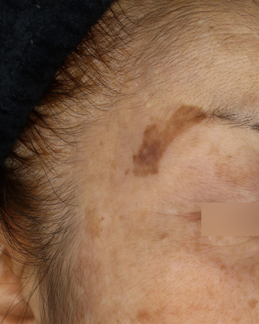
施術前
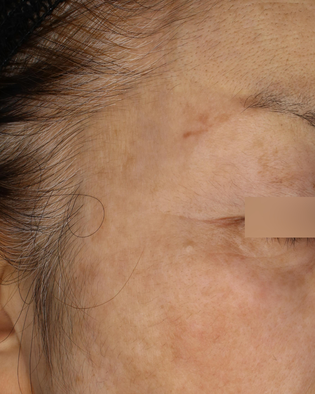
施術後
施術前
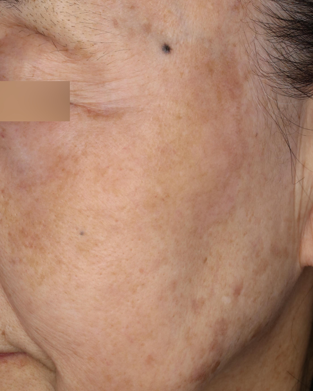
施術後
施術前
施術後
施術前
施術後
施術前
施術後
施術前
施術後
施術前
施術後
施術前
施術後
施術前
施術後
施術前
施術後
※症例写真は一例です。撮影条件、撮影時期、光の当たり方、肌の状態、治療経過には個人差があります。写真は治療効果を保証するものではなく、治療の適否は診察のうえで判断します。
TYPE 03|真皮型 —— 0.2ミリの下が中心にあるシミ
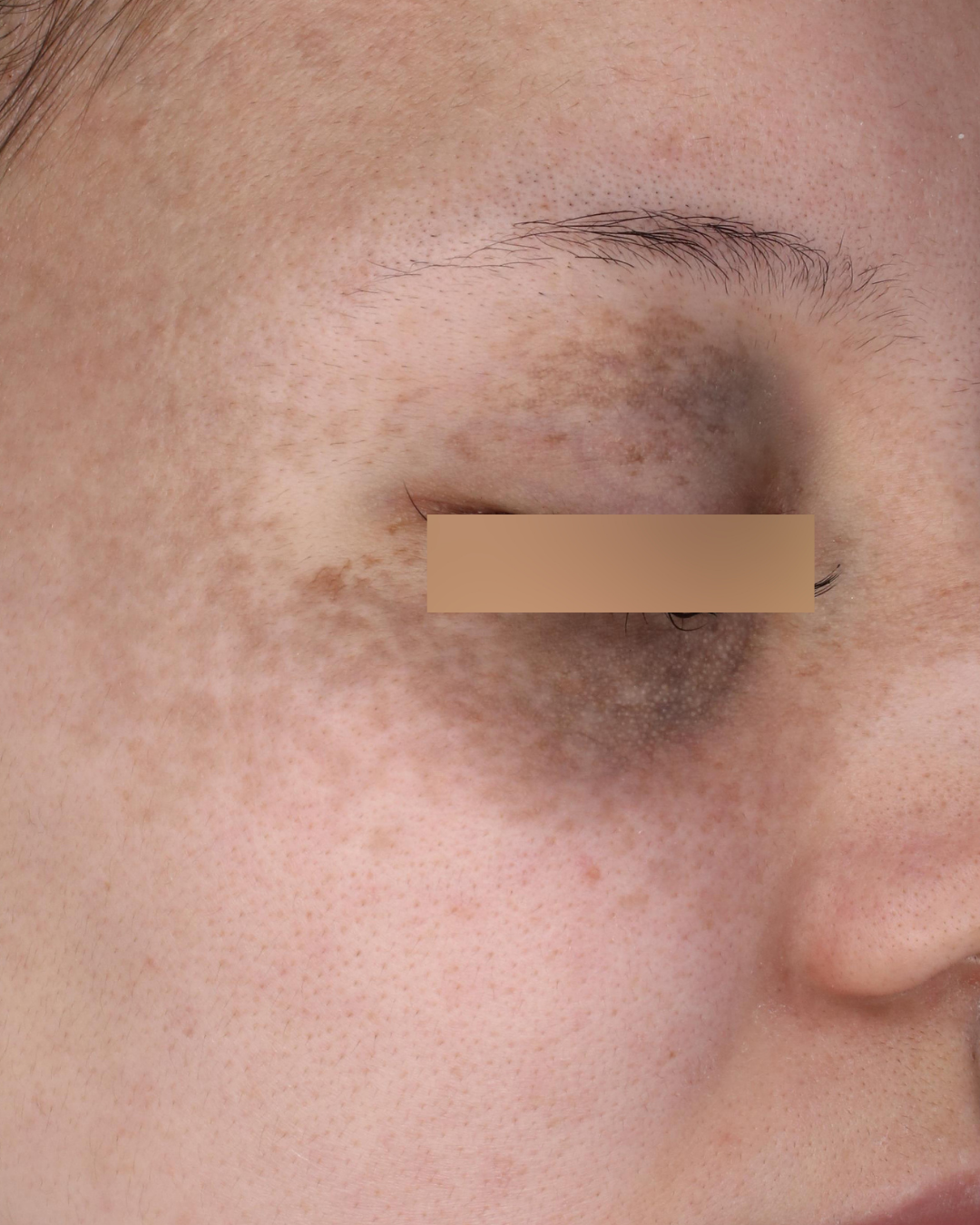
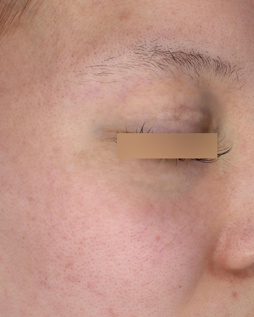
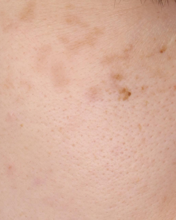
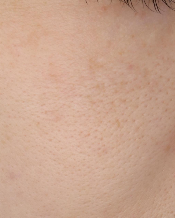
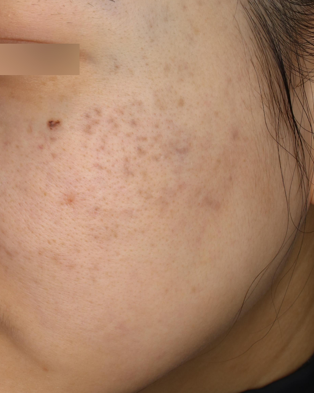
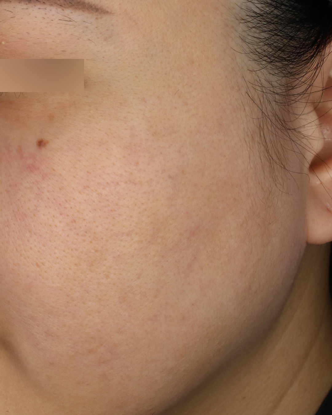
頬骨のあたりに左右対称に現れる灰色がかった斑。生まれつき、あるいは思春期以降に濃くなる青みのある色。ADM(後天性真皮メラノサイトーシス)や太田母斑は、色素の中心が真皮にある、根の深いタイプです。
真皮にあるこれらの深い色素には、同じ755nmでも、深部までエネルギーを届けるための設定と回数の設計が必要です。表面のシミと同じ照射では、根に届きません。逆に、真皮型を表皮のシミと見誤って浅い照射を重ねても、色は動かず、肌への負担だけが残ります。タイプを最初に読み違えないことが、このタイプでは特に重要です。
CASE PHOTOS — TYPE 03
真皮型の症例
施術前
施術後
施術前
施術後
施術前
施術後
※症例写真は一例です。撮影条件、撮影時期、光の当たり方、肌の状態、治療経過には個人差があります。写真は治療効果を保証するものではなく、治療の適否は診察のうえで判断します。
ここまでの3タイプ、ご自身のシミと重なるものはあったでしょうか。タイプは、診察で確かめられます。
開発の側から、この機器を知っているということ
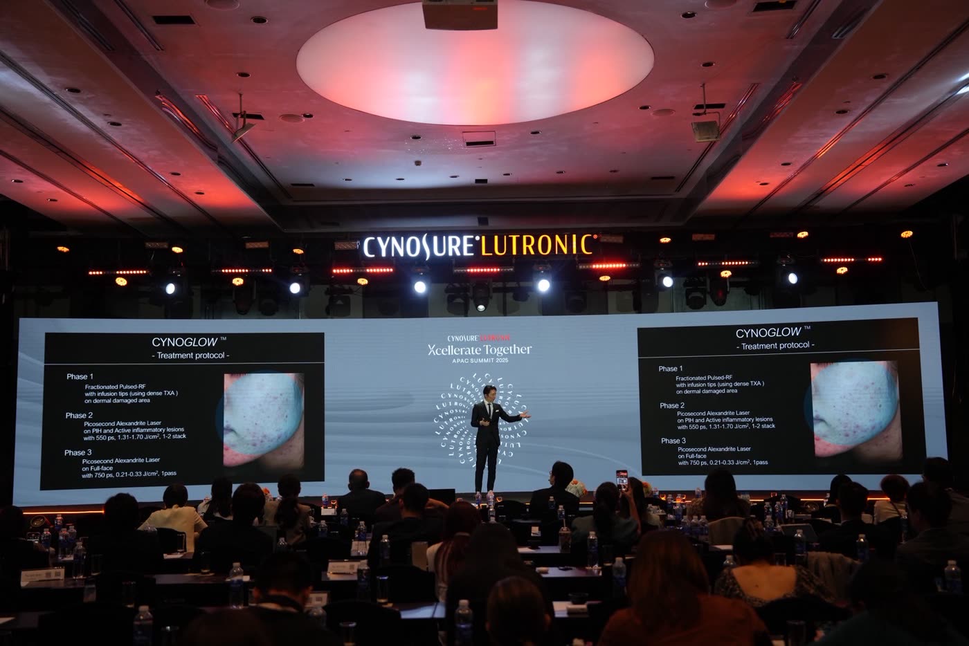
当院は前身機種PicoSureの時代から、色素治療に取り組んできました。私はPICOSURE proに開発段階から携わり、ローンチ後の調整と、照射方法の構築までを行ってきました。現在は、機器メーカーであるCynosure社のAdvisory Boardメンバーを日本人でただ一人務めています。当院のPICOSURE proは、その第1号機です。PICOSURE proに関する講演は全世界で90回以上、当院での照射は累計10,000例を超えています。
開発とローンチ後の調整を通して私が確かめてきたのは、この機器の得意なことよりも、どこからが過剰かという境目です。どの肌に、どの設定で、どこまでエネルギーを絞れば色素だけが壊れるのか。検証を重ねた分だけ、目の前の患者さまに「使わない設定」が増えていきます。
同じ機器でも、照射モード、出力、範囲、密度、治療間隔は一律ではありません。性能を知ることと、その性能を目の前の患者さまの肌に合わせて生かすことは、別の技術です。
レーザー後まで見据える、ヒルズフェイシャル®
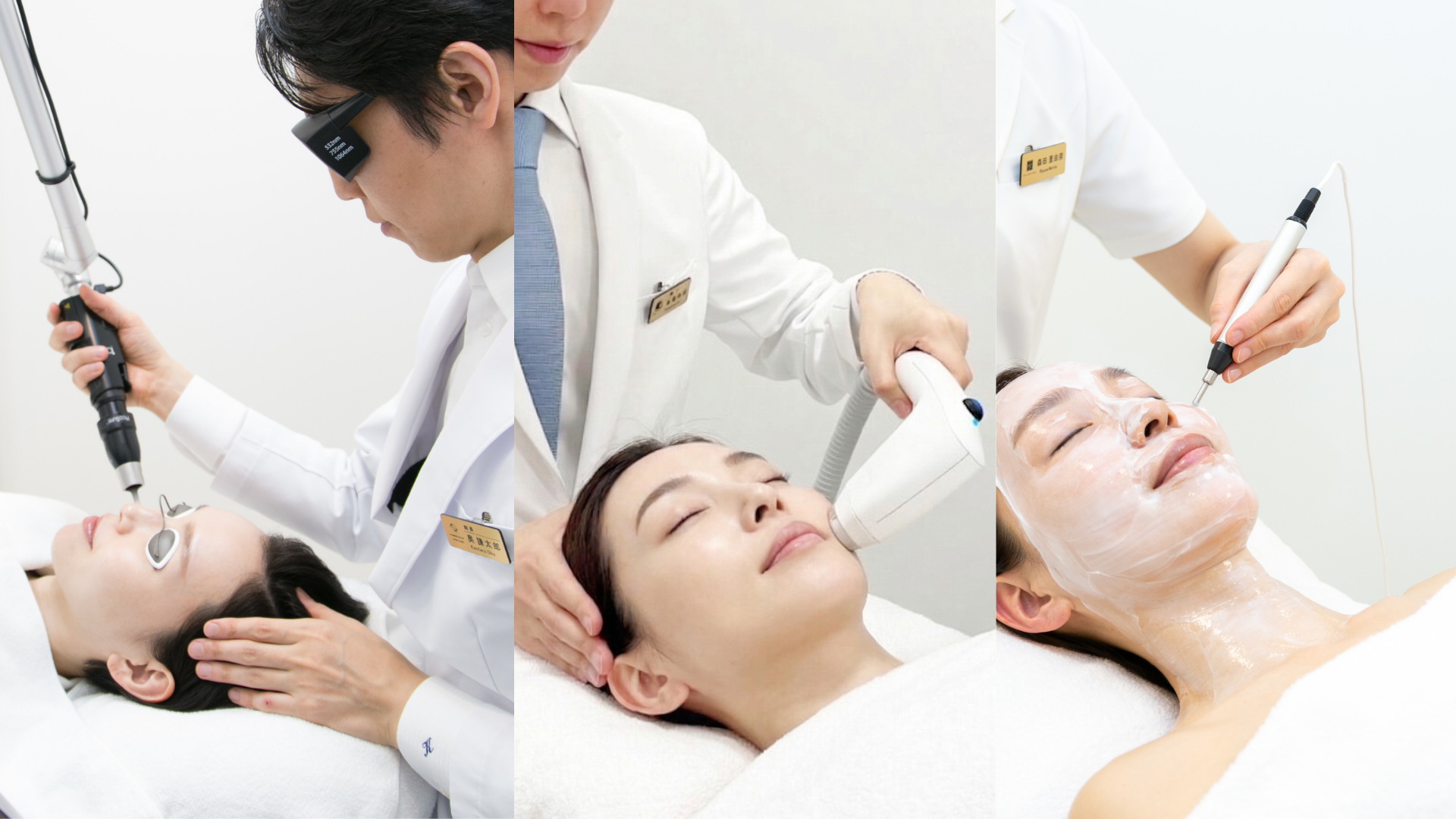
ヒルズフェイシャル®は、PICOSURE proとエレクトロポレーション「デルマシオ」を組み合わせる、全顔対象の治療プログラムです。細かな色むら、くすみ、質感まで含めて顔全体の印象を考え、照射後の導入ケアまでを一つの流れとして設計しています。
赤み、毛穴、ハリ、肌のゆるみなど複数のお悩みをあわせて考える場合は、ソフウェーブ、ダーマV、ポテンツァ Primeなどを組み合わせるヒルズフェイシャルプレミアム®を検討します。その方の肌状態に最適な機器をテーラーメイドで選び、熟練のテクニックで治療を行います。
初診で決めるのは、あなたのタイプです
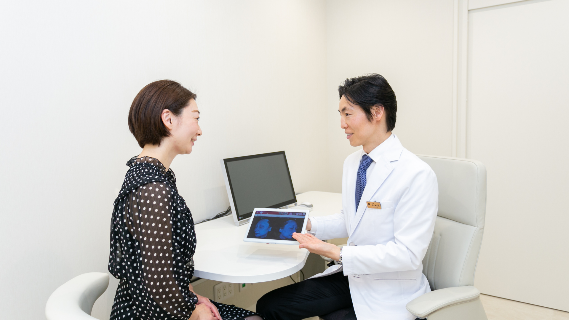
診察で行うのは、シミの種類と深さ、肌環境の診断です。あなたのシミがどのタイプにあたるのか。そのうえで、レーザーが適しているのか、先に肌を整えるべきなのか、見込む回数・ダウンタイム・費用はどのくらいかを、治療を始める前にお伝えします。
あなたのシミは、0.2ミリのどちら側にあるのか
あなたのシミは、0.2ミリのどちら側にあるのか。そして、肌の司令塔である線維芽細胞は、いま、どのモードで働いているのか。この二つを知るだけで、選ぶべき治療と、選ばなくていい治療が分かれます。
葉だけを刈るのか、根まで含めて設計するのか。眠った司令塔を起こしてから、エネルギーを届けるのか。診断すること。肌を整えること。必要なエネルギーを、必要な範囲に、必要な順番で届けること。治療後の経過まで見守ること。
シミを消すことではなく、これからの肌をどう整えていくか。私はその視点から、一人ひとりの治療を設計しています。
まずは、あなたのシミのタイプと、肌の状態を知ることから。
※治療の適応、回数、ダウンタイム、費用には個人差があります。赤み、腫れ、痛み、乾燥、色素沈着、色調変化、かさぶた、水疱、感染、瘢痕などが生じる可能性があります。治療内容は診察のうえで決定します。
よくある質問
自分がどのタイプかは、自分で分かりますか?
見た目だけで正確に判定することは難しく、複数のタイプが重なっていることも珍しくありません。診察では、シミの種類と深さ、肌環境、これまでの治療歴を合わせてタイプを確定します。ほくろ(色素性母斑)が混じっている場合は、診断を優先して治療方針を分けて判断します。
線維芽細胞のモードは、検査で分かりますか?
モードを直接測定する検査があるわけではありません。年齢、紫外線を浴びてきた期間、肌のハリや乾燥の状態、これまでの治療への反応などから、診察で総合的に評価します。光老化が進んだ肌では、CrossTALK® protocolで肌の土台を整えてからエネルギー治療へ進む順番をご提案することがあります。
0.2ミリは、実際に測定する数字ですか?
ここでいう0.2ミリは、表皮の厚さを目安に、色素が浅い層にあるのか深い層にあるのかを考えるための視点です。表皮の厚さは部位によって幅があります。診察では見た目だけでなく、シミの種類、肌環境、これまでの治療歴などを合わせて判断します。
一度取ったシミが、また出てくることはありますか?
あります。見えている色素を壊しても、色素を作る細胞の働きや、紫外線・摩擦・炎症などの肌環境が変わっていなければ、同じ場所に色が戻ることがあります。特に肝斑は刺激で悪化や再発を起こしやすいため、照射だけでなく外用・内服、スキンケアを含めた設計と、治療後の経過観察が大切です。
シミがあれば、すぐレーザーを受けたほうがよいですか?
すぐに照射をしない選択肢もあります。赤み、乾燥、炎症、摩擦の刺激が残っている場合は、紫外線対策やホームケア、外用・内服などを先に置くことがあります。肌の状態と治療の適応を診察で確認します。
PICOSURE proは、すべてのシミに向いていますか?
シミの種類、色素の深さ、肌の状態によって適する治療は変わります。PICOSURE proが適さない場合や、別の治療を先に検討する場合もあります。治療の適応、回数、ダウンタイムには個人差があります。
ヒルズフェイシャル®とプレミアム®は、何が違いますか?
ヒルズフェイシャル®はPICOSURE proとデルマシオを組み合わせる全顔対象のプログラムです。赤み、毛穴、ハリ、肌のゆるみなど複数のお悩みをあわせて考える場合は、ソフウェーブなどを組み合わせるプレミアム®を検討します。必要な治療だけを診察で選びます。
費用はどのくらいかかりますか?
公式料金の目安は、PICOSURE pro全顔が1回82,500円、ヒルズフェイシャル®全顔が1回115,500円です(自由診療・保険適用外)。シミの種類や範囲、組み合わせる治療によって変わるため、診察で治療計画とあわせてお見積りをお伝えします。詳しくは記事末尾の治療情報をご覧ください。
シミ・肝斑・毛穴など、肌に現れた悩みに向き合うとき、私が大切にしているのは、症状だけでなく、その症状が生じた肌の状態まで見極めることです。
レーザー・高周波・超音波などの機器は、同じ設定で照射しても、結果は一人ひとり異なります。その差を生むのは、肌が熱を受け止め、治療に応えられる状態にあるかどうか。だからこそ、必要に応じて肌が応えられる状態を整えながら、適切な治療を、適切な順序でご提案します。
この考えを治療設計の起点に据え、「Thermal Thread Technique®」として体系化し、2024年に『Lasers in Surgery and Medicine』誌へ発表しました。同論文は同誌のTop Viewed・Top Cited Articleに選出されています。高周波照射法「Mono-Bi CrossLIFT Technique」および、注入治療と医療機器を組み合わせる「CrossTALK® protocol」も『Journal of Cosmetic Dermatology』誌に発表しています。
米国レーザー医学会(ASLMS)専門医、医療法人社団光悠会理事長。Cynosure社のアドバイザリーボードには日本から唯一参加し、PicoSure Proの開発段階から携わり、第1号機による症例報告も行ってきました。世界最大のレーザー医学会ASLMSやIMCAS ASIA、KOREA DERMAなどの国際学会で招待講演を行い、国際的な医師教育プログラム「GMI」を主宰。海外の医師の指導・育成にも取り組んでいます。
これまでの治療で思うような手応えを得られなかった方にも、なぜその肌にその症状が起きているのかを見極め、必要な治療の順序からご提案します。肌を診て、治療を設計し、経過に合わせて組み立てを更新する。そこまでを含めて、肌治療だと考えています。詳しいプロフィール →
TREATMENT INFORMATION
治療について
- 施術名
- PICOSURE pro(ピコシュアプロ)全顔/ヒルズフェイシャル®(PICOSURE pro+デルマシオ)全顔/ヒルズフェイシャル®プレミアム(ソフウェーブ+PICOSURE pro等+デルマシオ)全顔/CrossTALK® protocol(スキンバイブ+ソフウェーブ等)
- 対象範囲
- PICOSURE pro全顔、ヒルズフェイシャル®全顔、ヒルズフェイシャル®プレミアム全顔。適応や使用機器の組み合わせは肌状態により異なります。
- 使用量・部位
- 全顔を基本としますが、照射モード、出力、範囲、密度、導入内容は診察で個別に決定します。本記事には施術前後の症例写真を掲載しています。
- 診療区分
- 自由診療(保険適用外)
- 公式料金の目安
-
PICOSURE pro
全顔 1回 82,500円
ヒルズフェイシャル®
全顔 1回 110,000円
ヒルズフェイシャル®プレミアム(HFP)
全顔 1回 280,000円
中顔面 1回 170,000円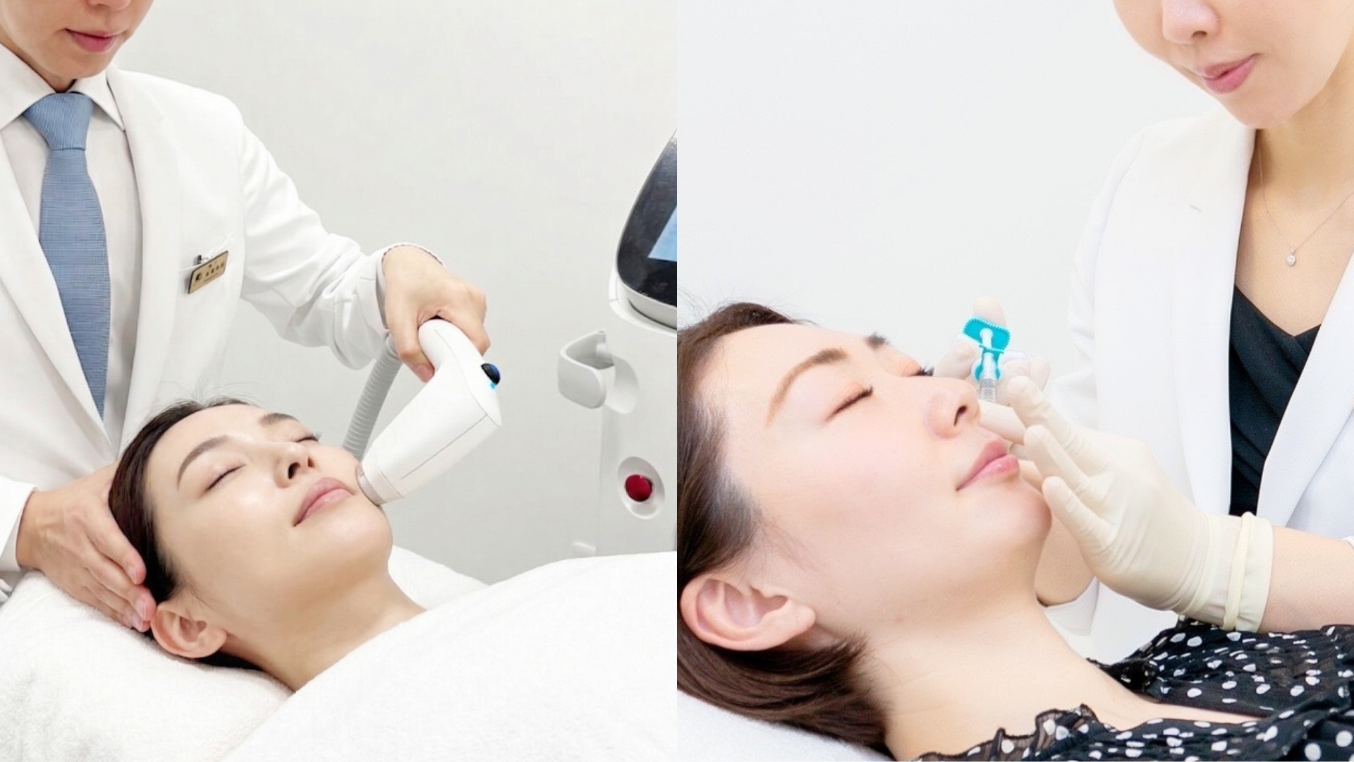
CrossTALK® protocol
全顔 1回 500,000円
中顔面 1回 270,000円上記は公式案内に掲載されている料金の目安です(税込・2026年10月時点。ヒルズフェイシャル®・プレミアムは2026年10月1日改定)。ピコシュアプロの表面麻酔代、ヒルズフェイシャル®プレミアムの麻酔代は別途かかります。CrossTALK® protocolは、麻酔代・内服代・針代などを含みます。診察・診断料は施術費用とは別に発生します(初めてご来院の方11,000円、前回のご来院から24か月以内の方1,100円、25か月以上の方11,000円)。料金や内容は変更される場合があります。
- 主な副作用・リスク
- PICOSURE proでは、皮膚不快感、紅斑、腫脹、点状出血、水疱形成、痂皮形成、内出血、色素沈着、膿疱、熱傷、白斑、色調の悪化、瘢痕、感染、アレルギー反応などが生じる可能性があります。施術後に赤みが残ることや、シミの部分が一時的に濃く見えることがあります。デルマシオでは紅斑、腫脹、アレルギー反応など、ソフウェーブでは熱感、痛み、赤み、腫れ、乾燥、まれに熱傷やしびれなどが生じる可能性があります。適応、回数、ダウンタイムには個人差があります。
- 未承認医療機器に関する事項
- PicoSure Proは国内承認医療機器です(承認番号 30500BZX00079000/一般的名称:アレキサンドライトレーザ/使用目的又は効果:体表面の表在性及び深在性良性色素性病変の治療、又は外傷性並びに入墨による刺青の蒸散及び除去)。ニキビ・ニキビ痕・毛穴・小じわ・肌質・肝斑を目的とする照射は、承認された使用目的の範囲外の適応外使用です。ソフウェーブは国内承認医療機器です(承認番号 30700BZX00082000/一般的名称:皮膚引締め用超音波照射器/承認された使用目的:中等度又は重度の顔面及び頚部のしわの改善)。引き上げ(たるみ)、毛穴、肌質、ニキビ痕、肝斑などを目的とする使用は、承認された使用目的の範囲外の適応外使用です。【デルマシオ(DERMATION)】本治療に使用するデルマシオは、日本国内において医薬品医療機器等法上の承認を得ていない未承認医療機器です。国内代理店であるNIOMA株式会社より入手しています。エレクトロポレーションによる美容成分の経皮導入を目的として国内で承認を受けた医療機器は、当院では確認できていません。製造元のある欧州では美容施術用の機器として販売されています。医療機器としての承認・認証の状況は確認中です。主なリスクとして、紅斑、腫脹、アレルギー反応等が報告されています。スキンバイブ by ジュビダームビスタ®は国内承認医療機器です(承認番号 30200BZX00389A01)。使用目的は、成人の顔面および頸部における小じわ等の表面のへこみ修正と皮膚状態(保水性、弾力性等)の改善で、眼瞼・口唇・隆鼻術における使用は適応に含まれません。治療内容は診察時に説明します。
PicoSure Proの公式案内 シミ治療の公式案内 ヒルズフェイシャル®の公式案内 CrossTALK® protocolの詳細(患者様ストーリー)
CLINIC INFORMATION
ヒルズグレイスクリニック
アクセスの詳細を見る →
肌を整える時間を、診察の外にも。

ENERGY DEVICE FRONTIER
Key points
Treatment for dark spots begins not with the name of a device, but with reading at what depth the pigment lies in relation to the epidermis, which is roughly 0.2 mm thick. In this article, I divide dark spots by depth into three types, “epidermal”, “mixed” and “dermal”, and introduce the approach to treatment for each type alongside 17 cases. The other key is the condition of fibroblasts, the skin’s command centre. Because cells that have dropped into a “low-activity mode” can be restored, our clinic has adopted a design in which the CrossTALK® protocol returns fibroblasts to regenerative mode before energy-based treatment is performed, and we have reported its efficacy and safety in an international journal. The type of your dark spots, and the condition of your skin. Knowing these two things is the most direct route to skin in which spots are less likely to recur.
Indications, number of sessions, downtime, cost and how treatment feels vary between individuals. Treatment is decided after an examination.
The epidermis is roughly 0.2 mm thick. Even with spots that look the same shade of brown, depending on whether the pigment lies shallower or deeper than this 0.2 mm, the treatment to choose is a different one. A laser treatment suited to shallow spots does not reach deep spots. Applying a treatment suited to deep spots to shallow spots may leave nothing but a burden on the skin.
Information about treatment for dark spots is everywhere. Precisely because we live in an age where it seems you can choose treatment by the name of a device, the first thing I do in an examination is not to choose a device. It is to read what is happening in this particular skin, and at what depth.
Even when they look similar, dark spots are not the same
Solar lentigines with well-defined edges. Fine freckles. Melasma spreading symmetrically on both sides. ADM in the dermis. Pigmentation left behind after inflammation.
In the examination, I check not only the type of pigment but also redness, dryness, any tendency to inflammation, habits that cause friction, opportunities for ultraviolet exposure and previous treatment history. This is because the skin’s response to laser treatment depends as much on the surrounding skin environment as on the pigment itself.
That is why there is sometimes a decision not to treat straight away. Hurrying to erase one conspicuous spot is not necessarily what is best for that person’s skin five years from now. Treatment for dark spots includes the decision not to apply the laser.
Which type are your dark spots?
The first thing I read in the examination is the depth of the pigment. Does the pigment lie above the roughly 0.2 mm line, overlap above and below it, or lie mainly below it? In this article, I discuss this by dividing it into three types.
Type Map — 0.2mm
Your dark spots: which type?
Solar lentigines, seborrhoeic keratosis, ephelides, etc.
Solar lentigines or ephelides coexisting with melasma, etc.
ADM, nevus of Ota, etc.
TYPE 01, the epidermal type, is a spot in which the pigment lies only above 0.2 mm. Solar lentigines, seborrhoeic keratosis and ephelides (freckles) fall into this group. TYPE 02, the mixed type, is the type in which these epidermal spots overlap with melasma spreading symmetrically on both sides. The mixed type is the one I encounter most often in the actual consultation room. TYPE 03, the dermal type, is the type in which the centre of the pigment lies below 0.2 mm, typified by ADM (acquired dermal melanocytosis) and nevus of Ota.
A guide to telling them apart in the mirror
From left: illustrations of the epidermal, mixed and dermal types.
Well-defined brown spots appear separately, one here, one there — a tendency of the epidermal type.
Pigment with blurred edges appears to spread almost symmetrically over the cheekbones — a tendency of the mixed type with melasma.
The spots look grey or bluish rather than brown — a tendency of the dermal type.
The type cannot be determined accurately by appearance alone. In the examination, we confirm it by considering the kind of spot, its depth and the skin environment together. Moles (pigmented naevi) are considered separately from “dark spots”. Diagnosis comes first, and whether or not to treat is decided on that basis.
Dark spots are like weeds
If you cut the leaves visible above ground, the view changes that same day. But if the roots remain in the soil, the weed grows back from the same place. Dark spots have a very similar structure.
The visible brown is melanin (the pigment that gives skin its colour) accumulated in the epidermis. In terms of a weed, these are the leaves above ground. This part, above the 0.2 mm line, can be broken up with a laser.
The roots correspond to the excessive activity of melanocytes (cells that produce pigment), which keep producing pigment at the base of the epidermis, and to the skin environment that keeps stimulating them. Ultraviolet light, friction, inflammation, hormonal changes. As long as the roots keep working, even if the surface pigment is cleared, colour tends to return to the same place over time.
Most cases of “the spot that was supposed to be gone has come back” are a state in which only the leaves were cut and the roots were left as they were. The three types can also be read through this metaphor. Only leaves growing thickly: the epidermal type. Roots that keep working beneath the leaves: the mixed type. Roots lying deep down: the dermal type.
TYPE 01 | Epidermal type — spots only above the 0.2 mm line
Solar lentigines with well-defined edges. Finely scattered ephelides. Slightly raised seborrhoeic keratoses. In each of these, the pigment lies mainly in the epidermis; in terms of a weed, the leaves are growing thickly. Treatment of this type begins by breaking up the visible pigment with the minimum of energy.
PICOSURE pro is a 755 nm picosecond laser. I use its ability to deliver energy in a very short time both to target pigment and in treatments that consider the texture of the skin as a whole.
The most important thing in picosecond laser treatment is not the strength of the output. What matters is identifying the lower limit: how much energy is enough to break up only the pigment. This threshold is called the threshold fluence.
It may seem that the higher the energy, the better it will work. In reality, more energy than necessary makes the surrounding normal tissue react as well and raises the risk of post-inflammatory hyperpigmentation (where the stimulus of treatment leaves the colour darker instead). Break up only what needs breaking up, and firmly protect what surrounds it. This, in my view, is the essence of treating dark spots.
Selective Protection
Only the dark spots, precisely targeted.
25–31%: Chan JC et al. Lasers Surg Med 2016 / 2.67%: compiled by Kentaro Oku from our clinic’s cases
In clinical data, the rate of post-inflammatory hyperpigmentation after PICOSURE pro treatment was 2.67% — a figure I compiled myself from our clinic’s cases. Laser treatment with the minimum necessary energy, based on identifying the threshold fluence, underpins this figure. That this is a picosecond laser with high efficacy and safety in treating dark spots has been shown by both basic medical data and clinical data.
Dark spots are determined by their background
Even among epidermal-type spots, the treatment design changes depending on the condition of the skin behind them. The key is the fibroblasts working in the dermis. They produce collagen, control the activity of the cells that make pigment, and are even involved in regulating immunity and blood vessels. They are, so to speak, the skin’s command centre.
This command centre has different states. Healthy fibroblasts grip collagen and are firmly stretched. This physical “stretch” itself becomes a signal to “be active”, and the cells keep actively producing collagen — this is regenerative mode. When collagen is broken down by ageing or ultraviolet light, the cells lose what they were gripping, shrink, and their activity drops as if they were asleep — low-activity mode. As this progresses further, cell division stops and the cells switch to releasing substances that promote inflammation and breakdown — senescent mode (Fisher GJ et al. Arch Dermatol 2008, among others).
Fibroblast Status
The three modes of fibroblasts, the skin’s command centre
In young skin with epidermal-type spots, the fibroblasts are working in regenerative mode. Because the heat from energy devices works to further accelerate collagen synthesis in cells that are already awake, Hills Facial®, which combines PICOSURE pro with the electroporation device Dermation, can on its own address both pigment and texture.
On the other hand, in epidermal-type skin with advanced photo-ageing, the command centre has dropped into low-activity mode. Fibroblasts in low-activity mode have reduced responsiveness to energy itself, and do not fully wake up when given heat alone. On the contrary, when a large amount of energy is applied to skin that has not been prepared, the heat can act simply as a burden.
What matters is that, unlike senescent mode, low-activity mode can be reversed. This is the point at which treatment intervenes. For skin of this kind, our clinic first works on the fibroblasts with the CrossTALK® protocol.
PUBLISHED IN
The Efficacy and Safety of Combining Cross-Linked Hyaluronic Acid Filler VYC-12L and Energy-Based Devices for Facial Skin Quality Improvement in Asians
When cross-linked hyaluronic acid (hyaluronic acid given a structure that resists breakdown) is placed in the skin, the internal pressure of the dermis rises and shrunken fibroblasts are physically stretched. This rebuilds the lost “scaffold to grip” and delivers to the cells the signal to “begin regeneration”. Once the cells in low-activity mode have returned to regenerative mode and the environment is in order, laser energy is delivered. This two-step protocol was prospectively evaluated in 18 patients, and we have reported in an international journal improvement in all patients on evaluator assessment, with no serious adverse events. Effects and progress vary between individuals.
CASE PHOTOS — TYPE 01
Epidermal-type cases
Before treatment
After treatment
Before treatment
After treatment
Before treatment
After treatment
Before treatment
After treatment
*Case photos are examples only. Photographic conditions, timing, lighting, skin condition and treatment progress vary between individuals. The photos do not guarantee treatment results, and suitability for treatment is determined after an examination.
TYPE 02 | Mixed type — spots overlapping above and below the 0.2 mm line
This is the type I see most often in the actual consultation room. Behind well-defined solar lentigines or ephelides, symmetrical melasma spreads faintly. More than half of the cases presented in this article are also of the mixed type.
What makes the mixed type difficult is that laser treatment that works on epidermal spots can instead act as a stimulus to melasma. The roots of melasma lie not so much in the pigment itself as in the excessive activity of the melanocytes that keep producing pigment and in the skin environment that stimulates them. This is why melasma tends to worsen or recur with changes in physical condition or irritation of the skin, and a strong laser treatment applied with good intentions can end up stimulating the roots.
That is why treatment of the mixed type is not complete with laser alone. We combine a low-stimulus laser treatment design with topical and oral medication, ultraviolet protection and skincare that reduces friction, to calm the activity of the roots themselves. Treatment that cuts the leaves, and treatment that weakens the roots. Only when both are in place can we aim for a state in which recurrence is less likely.
What you can do at home is also part of the treatment that weakens the roots. Choose a sunscreen that protects even against visible light. Do not rub. Keep using topical treatments. Use oral medicines such as tranexamic acid for a set period. In the examination, I explain how to keep these up as well.
And for the roots of the mixed type, our clinic has one more answer. In the roots of melasma, it has been reported that senescent fibroblasts keep sending out the instruction to “make pigment” (Ali L, Al Niaimi F. Int J Dermatol. 2025). In terms of the command-centre modes discussed in the section on the epidermal type, this is a state in which cells in senescent mode keep sending out signals, surrounded by cells in low-activity mode. Cells in senescent mode themselves can no longer be restored. However, it is possible to return the surrounding cells in low-activity mode to regenerative mode and rebuild the dermal environment. That is the CrossTALK® protocol. Cross-linked hyaluronic acid first creates a scaffold that fibroblasts can grip, and energy is applied once the command centre’s function has been restored. It has also been suggested that when the command centre regains its function, this works towards calming the excessive instructions sent to the cells that make pigment.
To use the weed analogy, rather than continuing to cut the leaves while the soil stays hard, this treatment tills the soil first and then addresses the roots. Once the foundation is in order, there is no longer any need to raise the output of each session, and even in melasma, which tends to worsen with stimulation, energy can be used not as a burden but as a trigger for recovery.
CASE PHOTOS — TYPE 02
Mixed-type cases
For the mixed type including melasma, the treatment design changes from case to case. Complex cases that include pigmented naevi are also handled by separating the treatment plans after diagnosis.
Before
After
Before
After
Before
After
Before
After
Before
After
Before
After
Before
After
Before
After
Before
After
Before
After
*The case photos are examples only. Photographic conditions, timing, lighting, skin condition and the course of treatment vary between individuals. The photos do not guarantee treatment results, and suitability for treatment is determined after a medical examination.
TYPE 03 | Dermal type — pigmentation centred below the 0.2 mm line
Greyish patches that appear symmetrically on both sides around the cheekbones. A bluish colour that is present from birth or darkens after puberty. ADM (acquired dermal melanocytosis) and naevus of Ota are deep-rooted types in which the pigment is centred in the dermis.
For these deep pigments in the dermis, even at the same 755 nm, the settings and number of sessions need to be planned to deliver energy to the deeper layers. Treatment set as for surface spots will not reach the roots. Conversely, if a dermal type is mistaken for epidermal pigmentation and shallow treatments are repeated, the pigmentation does not change, and only the burden on the skin remains. With this type in particular, it is important not to misread the type at the outset.
CASE PHOTOS — TYPE 03
Dermal-type cases
Before
After
Before
After
Before
After
*The case photos are examples only. Photographic conditions, timing, lighting, skin condition and the course of treatment vary between individuals. The photos do not guarantee treatment results, and suitability for treatment is determined after a medical examination.
Did any of these three types match your own dark spots? Your type can be confirmed at a consultation.
Knowing this device from the development side
Our clinic has been working on pigmentation treatment since the days of the predecessor device, PicoSure. I was involved with PICOSURE pro from the development stage, through post-launch adjustments, to building the treatment methods. I am currently the only Japanese member of the Advisory Board of Cynosure, the device manufacturer. The PICOSURE pro at our clinic is the very first unit.Lectures on PICOSURE pro have been given more than 90 times worldwide, and our clinic has performed more than 10,000 treatments in total.
What I have confirmed through development and post-launch adjustment is not so much what this device does well as the boundary beyond which it becomes excessive. For which skin, at which settings, and how far must the energy be narrowed so that only the pigment is broken down? The more verification I carry out, the more “settings not to use” I have for the patient in front of me.
Even with the same device, the treatment mode, power, area, density and treatment interval are not uniform. Knowing a device’s performance and putting that performance to use for the skin of the patient in front of you are separate skills.
Hills Facial®, planned with what comes after the laser in mind
Hills Facial® is a full-face treatment programme that combines PICOSURE pro with the electroporation device “Dermation”. It considers the overall impression of the face, including fine uneven pigmentation, dullness and texture, and is designed as a single process that continues through to infusion care after the laser treatment.
When several concerns such as redness, pores, firmness and skin laxity are considered together, we consider Hills Facial Premium®, which combines Sofwave, DermaV, Potenza Prime and other devices. We select the most suitable devices for each person’s skin condition on a tailor-made basis and perform the treatment with skilled technique.
What is decided at your first visit is your type
At the consultation, we diagnose the type and depth of the dark spots and the skin environment: which type your dark spots fall into. On that basis, whether laser is suitable or the skin should be prepared first, and the expected number of sessions, downtime and cost — we explain these to you before treatment begins.
Which side of 0.2 mm are your dark spots on?
Which side of 0.2 mm are your dark spots on? And in which mode are your fibroblasts, the command centre of the skin, working right now? Simply knowing these two things separates the treatments you should choose from those you need not choose.
Do we cut back only the leaves, or plan to include the roots? Do we wake the dormant command centre before delivering energy? Diagnosing. Preparing the skin. Delivering the energy that is needed, to the area that needs it, in the order it is needed. Watching over the course after treatment.
Not erasing dark spots, but how to care for your skin from here on. It is from that perspective that I design each person’s treatment.
Begin by learning your dark-spot type and the condition of your skin.
Book a consultation for dark spots and melasma
*Suitability for treatment, number of sessions, downtime and cost vary between individuals. Redness, swelling, pain, dryness, hyperpigmentation, changes in skin tone, scabbing, blistering, infection, scarring and other effects may occur. The treatment is decided after a medical examination.
Frequently asked questions
Can I tell which type I am myself?
It is difficult to determine the type accurately from appearance alone, and it is not unusual for several types to overlap. At the consultation, we confirm the type by considering the type and depth of the dark spots, the skin environment and your treatment history together. If moles (pigmented naevi) are mixed in, diagnosis takes priority and the treatment plan for them is decided separately.
Can the mode of my fibroblasts be determined by a test?
There is no test that measures the mode directly. We assess it comprehensively at the consultation, based on age, how long the skin has been exposed to UV, the skin’s firmness and dryness, its response to previous treatments, and so on. For skin with advanced photo-ageing, we may suggest preparing the foundation of the skin with the CrossTALK® protocol before moving on to energy-based treatment.
Is 0.2 mm a figure that is actually measured?
The 0.2 mm referred to here is a perspective for considering whether the pigment lies in a shallow or a deep layer, using the thickness of the epidermis as a guide. The thickness of the epidermis varies by area of the body. At the consultation, we judge not only by appearance but also by the type of dark spot, the skin environment, your treatment history and other factors.
Can a dark spot that has been removed come back?
Yes. Even if the visible pigment is broken down, the pigmentation may return in the same place if the activity of the pigment-producing cells and the skin environment, such as UV, friction and inflammation, have not changed. Melasma in particular is prone to worsening or recurring with irritation, so it is important to plan care that includes not only laser treatment but also topical and oral medication and skincare, and to follow the course after treatment.
If I have dark spots, should I have laser treatment straight away?
Not having laser treatment straight away is also an option. If redness, dryness, inflammation or irritation from friction persists, UV protection, home care, topical and oral medication or other measures may come first. We check the condition of your skin and your suitability for treatment at a consultation.
Is PICOSURE pro suitable for all dark spots?
The suitable treatment depends on the type of dark spot, the depth of the pigment and the condition of the skin. In some cases PICOSURE pro is not suitable, or another treatment may be considered first. Suitability for treatment, number of sessions and downtime vary between individuals.
What is the difference between Hills Facial® and Premium®?
Hills Facial® is a full-face programme that combines PICOSURE pro with Dermation. When several concerns such as redness, pores, firmness and skin laxity are considered together, we consider Premium®, which combines Sofwave and other devices. Only the treatments that are needed are selected at the consultation.
How much does it cost?
The standard fees are ¥82,500 per session for PICOSURE pro full face and ¥115,500 per session for Hills Facial® full face (private, self-funded care, not covered by health insurance). Because the fee varies with the type and extent of the dark spots and the treatments combined, we give you an estimate together with your treatment plan at the consultation. For details, please see the treatment information at the end of this article.
When addressing concerns that appear on the skin, such as dark spots, melasma and pores, what I value is looking not only at the symptoms but also at the condition of the skin in which those symptoms arose.
With devices such as lasers, radiofrequency and ultrasound, results differ from person to person even at the same settings. What creates that difference is whether the skin is in a state to receive heat and respond to treatment. That is why, where necessary, I prepare the skin so that it can respond, while suggesting appropriate treatments in the appropriate order.
Placing this idea at the starting point of treatment design, I systematised it as the “Thermal Thread Technique®” and published it in “Lasers in Surgery and Medicine” in 2024. The paper was selected as a Top Viewed and Top Cited Article in the journal. I have also published the radiofrequency technique “Mono-Bi CrossLIFT Technique” and the “CrossTALK® protocol”, which combines injectable treatment with medical devices, in the “Journal of Cosmetic Dermatology”.
Specialist of the American Society for Laser Medicine and Surgery (ASLMS); Chair of the Board, Koyukai Medical Corporation. As the only participant from Japan on Cynosure’s advisory board, I have been involved with PicoSure Pro since its development stage and have also reported cases treated with the first unit. I have given invited lectures at international congresses such as ASLMS, the world’s largest laser medicine society, IMCAS ASIA and KOREA DERMA, and I lead “GMI”, an international physician education programme. I am also engaged in teaching and training doctors from overseas.
For those who have not had the response they hoped for from previous treatments, too, I look into why that symptom is occurring in that skin and suggest treatment starting from the order in which it is needed. Examining the skin, designing the treatment, and updating the plan as the course progresses: I consider all of this to be skin treatment. Full profile →
TREATMENT INFORMATION
About the treatment
- Treatment name
- PICOSURE pro full face / Hills Facial® (PICOSURE pro + Dermation) full face / Hills Facial® Premium (Sofwave + PICOSURE pro, etc. + Dermation) full face / CrossTALK® protocol (SKINVIVE + Sofwave, etc.)
- Treatment area
- PICOSURE pro full face, Hills Facial® full face, Hills Facial® Premium full face. Suitability and the combination of devices used depend on the condition of the skin.
- Amount and area
- Treatment is basically full face, but the treatment mode, power, area, density and infusion content are decided individually at the consultation. This article includes before-and-after case photos.
- Category of care
- Private, self-funded care (not covered by health insurance)
- Standard fees
-
PICOSURE pro
Full face, 1 session ¥82,500
Hills Facial®
Full face, 1 session ¥110,000
Hills Facial® Premium (HFP)
Full face, 1 session ¥280,000
Mid-face, 1 session ¥170,000
CrossTALK® protocol
Full face, 1 session ¥500,000
Mid-face, 1 session ¥270,000The above are standard fees as listed in the official information (tax included, as of October 2026; Hills Facial® Premium fees revised on 1 October 2026). The topical anaesthetic fee for PicoSure Pro and the anaesthesia fee for Hills Facial® Premium are charged separately. The CrossTALK® protocol fee includes anaesthesia, oral medication, needles and similar costs. Consultation and diagnosis fees are charged separately from treatment costs (¥11,000 for first-time patients, ¥1,100 for patients within 24 months of their previous visit, ¥11,000 for patients 25 months or more after their previous visit). Fees and details may change.
- Main side effects and risks
- With PICOSURE pro, skin discomfort, erythema, swelling, petechiae, blistering, crusting, bruising, hyperpigmentation, pustules, burns, leukoderma, worsening of skin tone, scarring, infection, allergic reactions and other effects may occur. Redness may remain after treatment, and the dark spots may temporarily look darker. With Dermation, erythema, swelling, allergic reactions and other effects may occur; with Sofwave, a sensation of heat, pain, redness, swelling, dryness and, rarely, burns or numbness may occur. Suitability, number of sessions and downtime vary between individuals.
- Unapproved medical devices
- PicoSure Pro is a medical device approved in Japan (approval number 30500BZX00079000; generic name: alexandrite laser; intended use or effect: treatment of superficial and deep benign pigmented lesions on the body surface, or vaporisation and removal of traumatic and decorative tattoos). Treatment aimed at acne, acne scars, pores, fine wrinkles, skin quality or melasma is off-label use outside the scope of the approved intended use. Sofwave is a medical device approved in Japan (approval number 30700BZX00082000; generic name: ultrasound device for skin tightening; approved intended use: improvement of moderate or severe facial and neck wrinkles). Use aimed at lifting (sagging), pores, skin quality, acne scars, melasma and similar concerns is off-label use outside the scope of the approved intended use. [Dermation (DERMATION)] The Dermation device used in this treatment is an unapproved medical device that has not received approval under the PMD Act in Japan. It is obtained from NIOMA Co., Ltd., the domestic distributor. Our clinic has not been able to confirm any medical device approved in Japan for the transdermal delivery of cosmetic ingredients by electroporation. In Europe, where the manufacturer is based, it is sold as a device for aesthetic treatments. Its approval or certification status as a medical device is being confirmed. The main reported risks include erythema, swelling and allergic reactions. SKINVIVE by JUVÉDERM VISTA® is a medical device approved in Japan (approval number 30200BZX00389A01). Its intended use is the correction of surface depressions such as fine wrinkles on the face and neck of adults and the improvement of skin condition (hydration, elasticity, etc.); use on the eyelids or lips, or in rhinoplasty, is not included in its indications. Details of the treatment are explained at the consultation.
Official information on PicoSure Pro Official information on dark-spot treatment Official information on Hills Facial® CrossTALK® protocol in detail (patient story)
CLINIC INFORMATION
Hills Grace Clinic
Access details →
Caring for your skin beyond the consultation room
HOME CARE
Online shop
Doctor-selected skincare and supplements, delivered to your home. For some products, a free online consultation is available before purchase.
CONSULTATION
Consult us at the clinic
Every person's skin is different. We look into what lies behind pigmentation and melasma, and suggest the care and the order of treatment your skin needs now.